Figure 2. Fundus photographs of
affected individuals. Fundus photographs of the right eye from
affected individual II:4 A:, affected individual II:6 B:
affected individual II:7 C: and the unaffected father
I:1 D: who carries the mutation heterozygously. The ages
at the time of investigation of individuals II:4, II:6, and II:7
were 36, 28, and 26, respectively. Fundus examination of the
affected individuals revealed typical features of retinitis
pigmentosa, bone spicule-like pigmentations were found in the
mid periphery of retina (indicated with arrows), and retinal
vessels were attenuated (arrowheads).
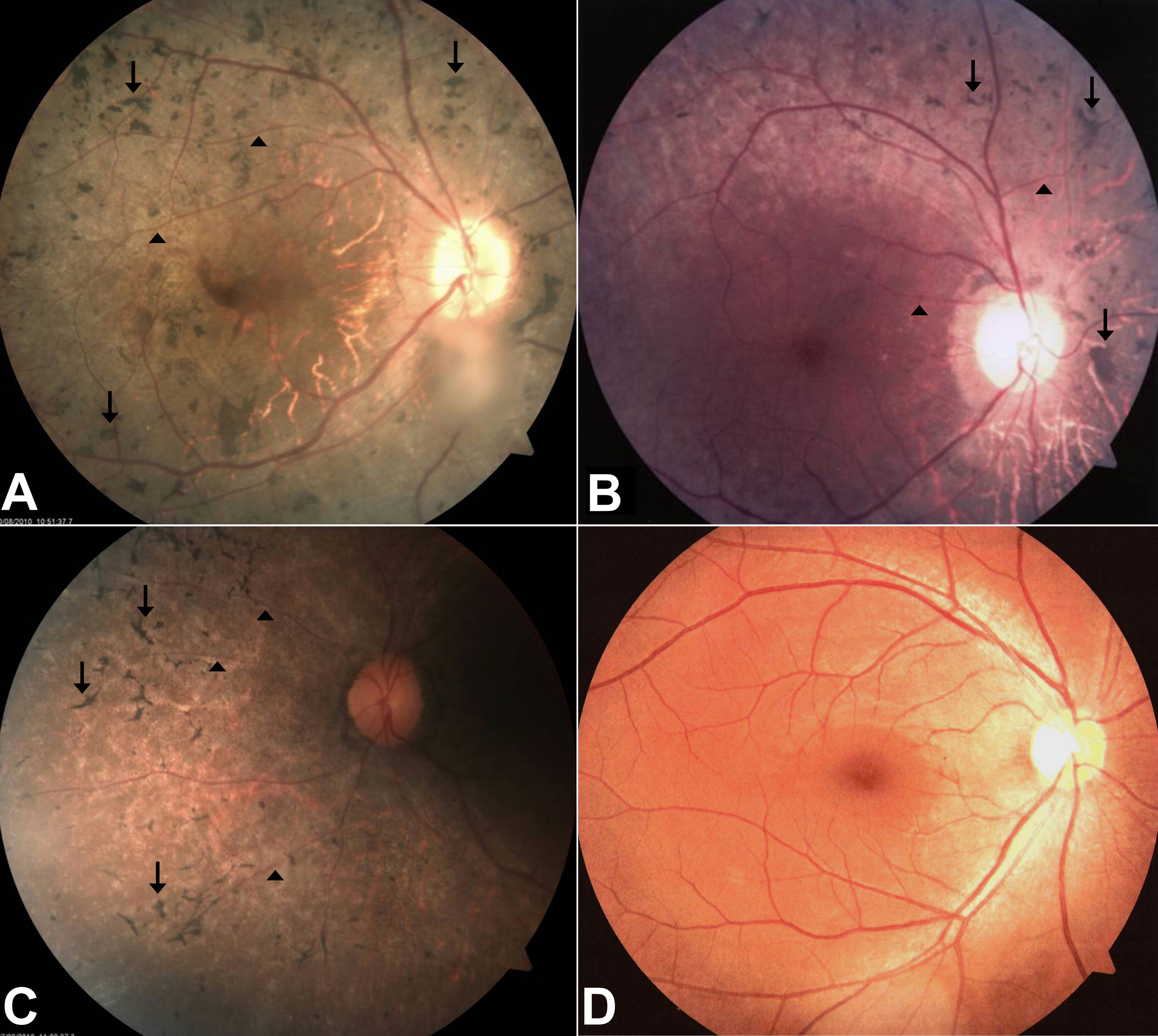
 Figure 2
of Siemiatkowska, Mol Vis 2012;
18:2411-2419.
Figure 2
of Siemiatkowska, Mol Vis 2012;
18:2411-2419.