Figure 5. Expression and distribution
of heat shock protein 70 in RF/6A cells after laser irradiation.
A-C: Unirradiated RF/6A cells. D-F:
RF/6A cells were treated with 700 mW laser irradiation and
followed by 18 h incubation. Cells were fluorescently labeled
with heat shock protein 70 (Hsp70; center, red channel) and the
nuclear marker diamidinophenylindole (left, blue channel). The
right is an overlay of the two channels. A strong induction of
Hsp70 was observed in cells treated with a laser power of 700
mW. The induced Hsp70 was present in both the cytoplasm and
nucleus. Data are from one of three separate experiments with
similar results.
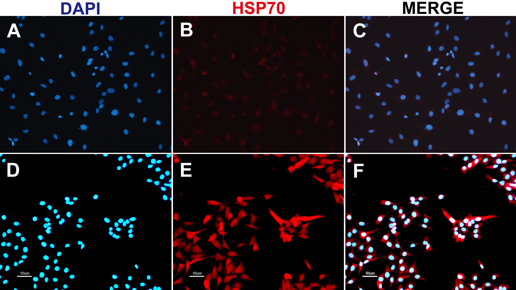
 Figure 5
of Du, Mol Vis 2012; 18:2380-2387.
Figure 5
of Du, Mol Vis 2012; 18:2380-2387.