Figure 2. Micrographs of RF/6A cells
treated with different laser powers. A: Unirradiated
RF/6A cells. B: RF/6A cells treated with 1,000 mW laser
irradiation. C: RF/6A cells treated with 1,100 mW laser
irradiation. Cells with apoptotic or necrotic morphology were
present in the 1,100 mW treated samples. Data are from one of
three separate experiments with similar results.
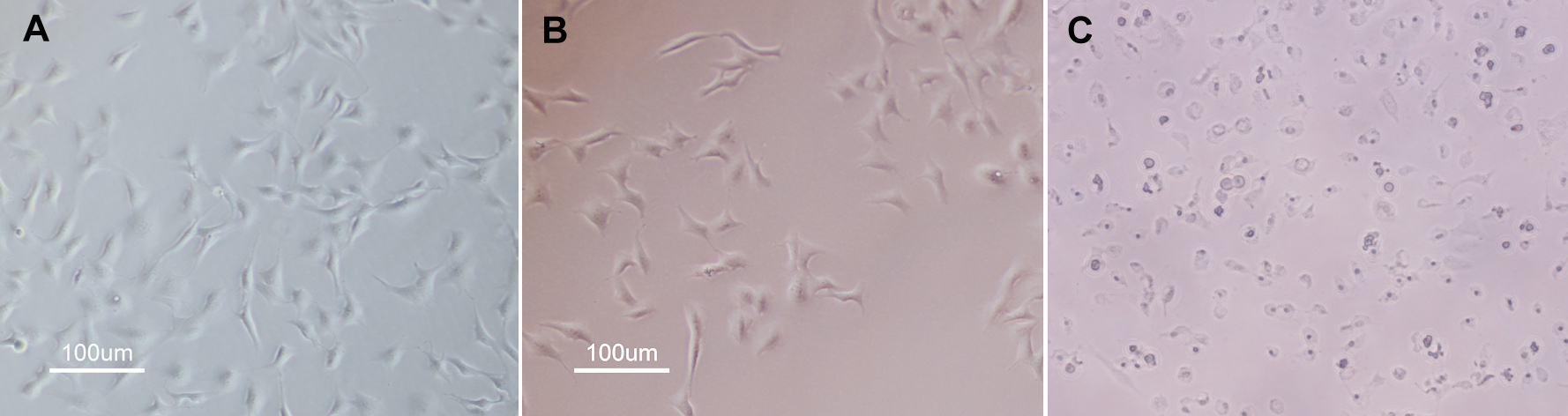
 Figure 2
of Du, Mol Vis 2012; 18:2380-2387.
Figure 2
of Du, Mol Vis 2012; 18:2380-2387.