Figure 3. Hyperglycemia promotes
CaMKll activation in RF/6A cells. A: RF/6A cells were
incubated for 96 h in a serum-free medium with 5.5 mM glucose
(NG), 30 mM glucose (HG), or HG plus 2-APB, and subjected to
western blotting analysis for CaMKll and p-CaMKll protein
levels. β-actin served as loading control. B: CaMKll and
p-CaMKll levels were quantified by densitometry analysis under
each treatment condition. Bars represented mean±SD from at least
three independent experiments with seven cells per treatment
group. * p<0.05 versus NG; # p<0.05 versus HG;
# p>0.05 versus NG.
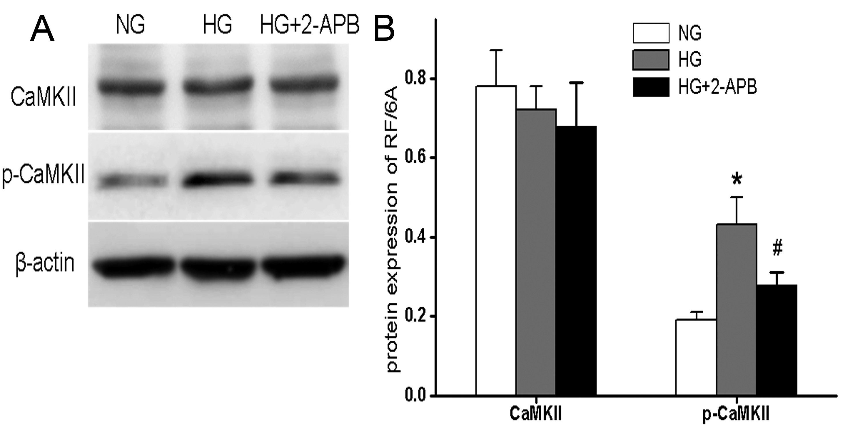
 Figure 3
of Li, Mol Vis 2012; 18:2371-2379.
Figure 3
of Li, Mol Vis 2012; 18:2371-2379.