Figure 5. Representative images from
immunohistochemistry 80 days after intravitreal injection of
plasmin and SF6 (group 1), plasmin (group 2), SF6
(group 3), or balanced salt solution (control group). There were
no significant differences in the retinal cell components
between eyes receiving plasmin and/or SF6 and
balanced salt solution (BSS). The scale bar represents 150 μm.
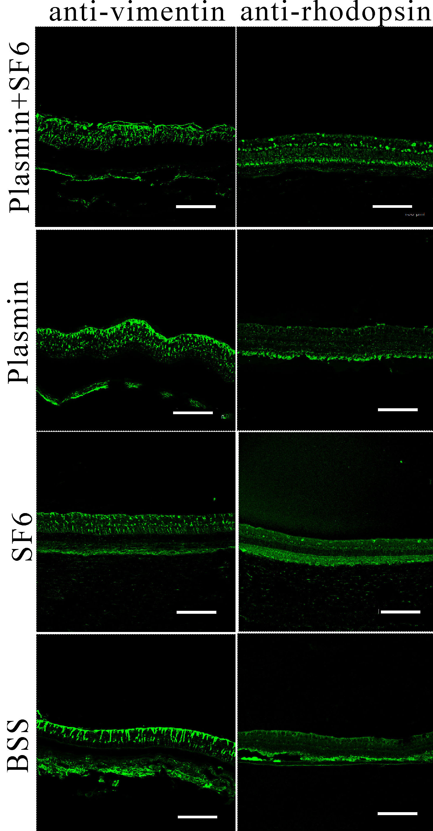
 Figure 5
of Wu, Mol Vis 2012; 18:2361-2370.
Figure 5
of Wu, Mol Vis 2012; 18:2361-2370.