Figure 7. RTL220 protects the degenerating retina from microglia/macrophages (MG/mφ) activities. A-C: Representative confocal photomicrographs of the retina show the preservation of photoreceptor cells and reduction in MG/mφ
after RTL220 treatment at P60. There is a significant reduction in MG/mφ migration and their presence in subretinal debris
space in RTL220-treated retinas (C). MG/mφ were stained with lba-1 antibody (red) and nuclei with diamidino-phenyl-indole (DAPI; blue). D-F: Representative images show immunofluorescent staining of Royal College of Surgeons (RCS) rat retinas with anti- monocyte
chemotactic protein-1 (MCP-1) antibodies (red) and nuclei with DAPI (blue). D: dystrophic retina at P30 shows MCP-1 expression around the ganglion cell layer; E: RTL101-treated retina. F: RTL220- treated retina shows a reduction in MCP-1 fluorescence; arrows indicate labeling of MCP-1 (red). Abbreviations:
OS - outer segments, ONL - outer nuclear layer, INL - inner nuclear layer, GCL - ganglion cell layer.
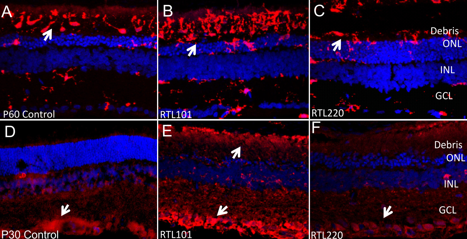
 Figure 7 of
Adamus, Mol Vis 2012; 18:2323-2337.
Figure 7 of
Adamus, Mol Vis 2012; 18:2323-2337.