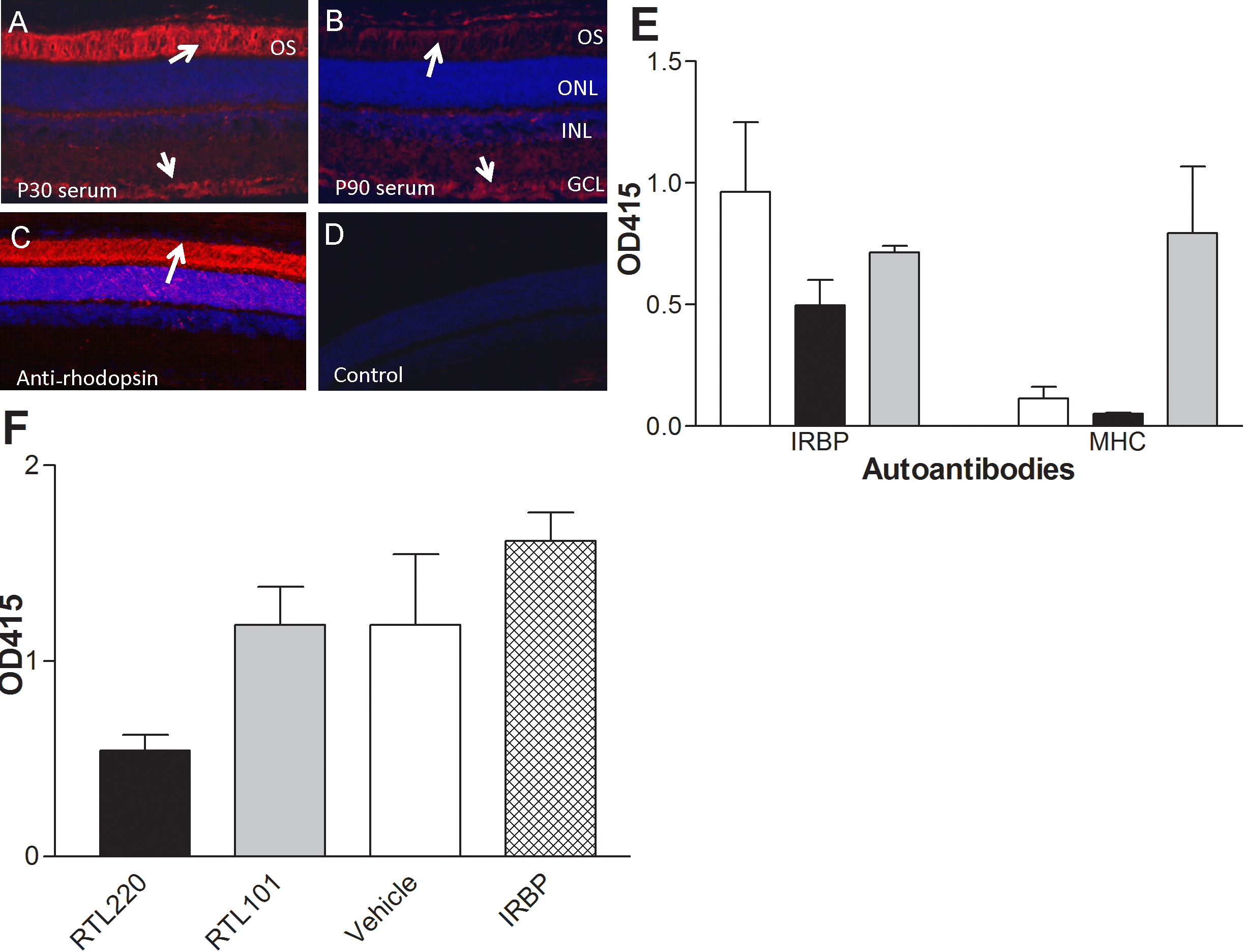
Figure 6. Immune responses in RCS
rats.. A-D: Confocal images demonstrate labeling of
non-dystrophic rat retina with autoimmune sera from untreated
dystrophic RCS rats that was collected at P30 and P90 (dilution
1:20). A: Strong labeling of outer and inner segments
was present at P30 (arrows), B: weaker immunolabeling of
ganglion cell layer and outer segments in rat retina with serum
antibodies collected at P90. C: Control staining of the
non-dystrophic rat retina with anti-rhodopsin mAb (R2–12N,
1:1000) that strongly immunolabeled the outer and inner segments
of photoreceptors. D: No labeling of retinal cells was
observed in control immunolabeling without primary antibodies. E,
F: RTL220 treatment inhibits autoantibody response
against interphotoreceptor retinoid binding protein (IRBP). E:
The bars represent autoantibody titers against IRBP1177–1191 and
β1α1 MHC polypeptide (part of RTL) in serum from P60 rats (n=5)
diluted 1:100 and measured by enzyme-linked immunosorbent assay
(ELISA). Note that RTL220 decreased autoantibodies against
IRBP1177–1191 peptide (black bars). Also rats produced
antibodies against MHC when treated with RTL101 (β1α1 chain of
MHC, gray bars). Vehicle treatment-white bar. F:
RTL220-treated significantly reduced autoantibody levels against
IRBP1177–1191 peptide (n=6, one-way ANOVA, p=0.0076) in sera
from rats at P90 measured by ELISA at 1:100 dilutions. OS -
outer segments, ONL - outer nuclear layer, INL - inner nuclear
layer, GCL - ganglion cell layer, AAb - autoantibodies, MHC -
major histocompatibility complex.
Adamus, Mol Vis 2012; 18:2323-2337. http://www.molvis.org/molvis/v18/a246
©2012 Molecular Vision http://www.molvis.org/molvis/
ISSN 1090-0535
 Figure 6
of Adamus, Mol Vis 2012; 18:2323-2337.
Figure 6
of Adamus, Mol Vis 2012; 18:2323-2337.