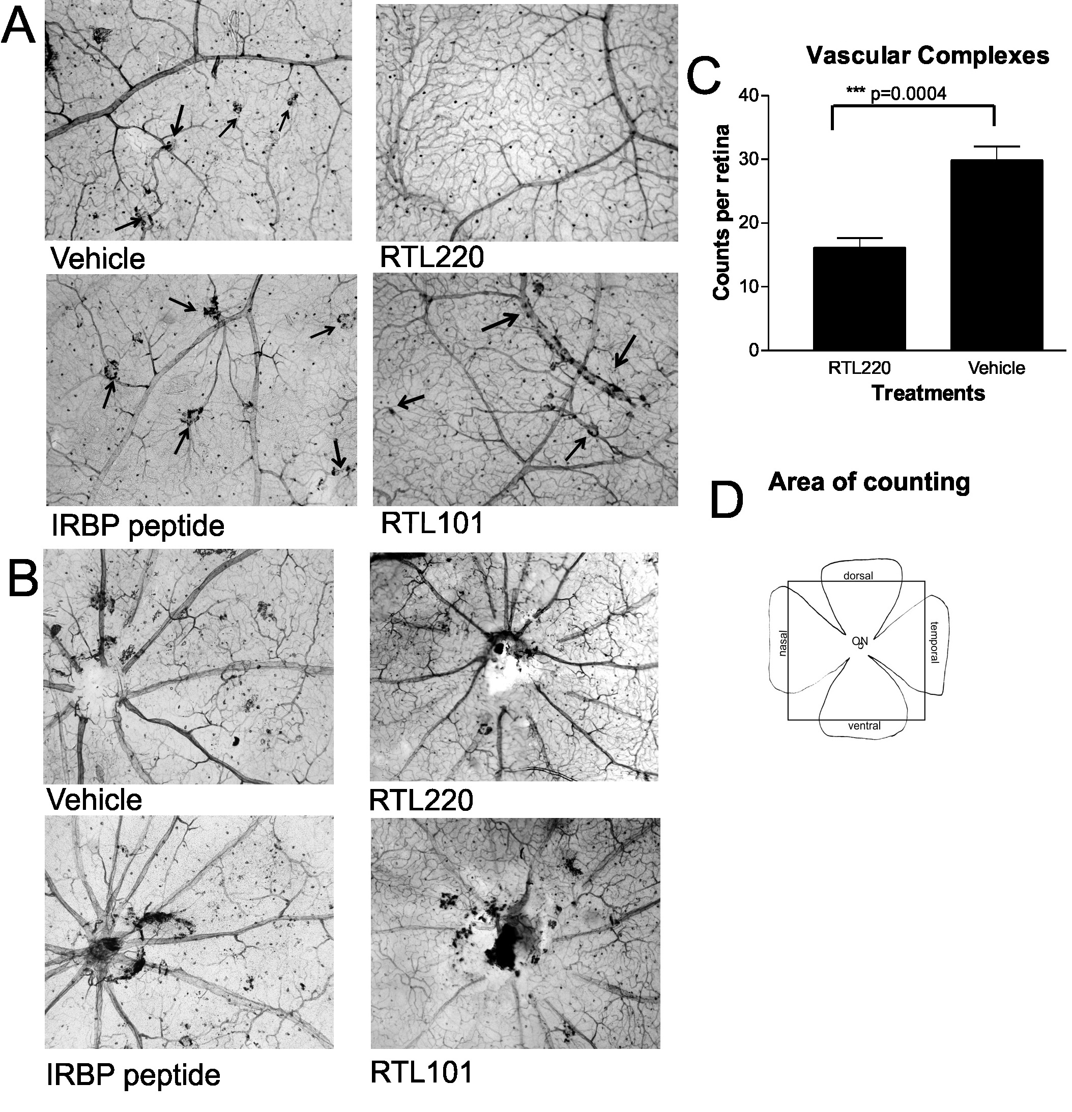
Figure 4. Representative vascular
pathology is shown for retinal whole mounts stained with
nicotinamide adenine dinucleotide phosphate (NADPH)-diaphorase
to identify abnormal vessels associated with retinal pigment
epithelium (RPE) cells (vascular complexes; arrows). A:
Photomicrographs show the vascular complexes from middle part of
the retina that were dramatically reduced in RTL220-treated rats
compared to control rats. RTL101 was not effective and its
effect was similar to vehicle- or plain interphotoreceptor
retinoid binding protein (IRBP) peptide-treated RCS rats. B:
Optic nerve head region view of RTL220 and control RTL101
treatment show the accumulation of pigmented cells around optic
disc. C: Bar graph represents the overall reduction in
vascular complexes in RTL220-treated retinas compared to
vehicle-treated retinas (n=6, p=0.0004). D: Scheme of
retinal flatmounts shows the area from which the abnormal
vessels were counted (box).
Adamus, Mol Vis 2012; 18:2323-2337. http://www.molvis.org/molvis/v18/a246
©2012 Molecular Vision http://www.molvis.org/molvis/
ISSN 1090-0535
 Figure 4
of Adamus, Mol Vis 2012; 18:2323-2337.
Figure 4
of Adamus, Mol Vis 2012; 18:2323-2337.