Figure 3. Typical angiography of
retinal whole mounts is shown for different Royal College of
Surgeons (RCS) rat treatment groups. A: Fluorescent
micrographs represent RTL220, RTL101, IRBP1177–1191 peptide and
vehicle-treated rats. The fluorescent pictures were taken from
middle retinas around optic disc regions in A (RTL220
and RTL101), showing protective effects of RTL220 on the
vasculature compared to the control rats presenting vascular
leakage and abnormal vessels (arrows). B: Bar graph show
the reduction in total counts of leaking vessels in
RTL220-treated rats compared to the vehicle-treated rats (n=6,
p=0.0018). C: Scheme of retinal flat mounts shows the
area from which the leaking vessels were counted (box).
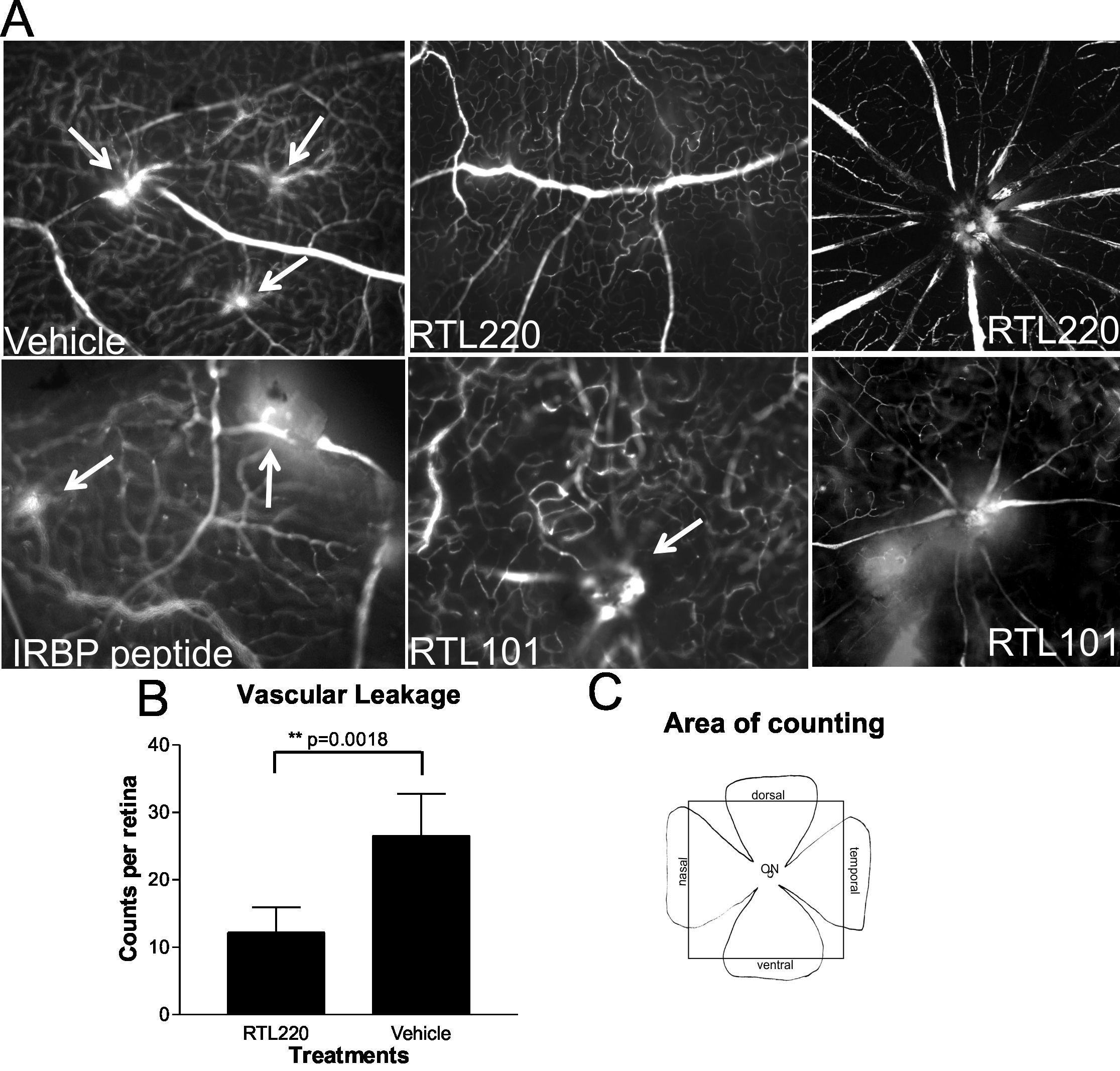
 Figure 3
of Adamus, Mol Vis 2012; 18:2323-2337.
Figure 3
of Adamus, Mol Vis 2012; 18:2323-2337.