Figure 1. Retinal protection by RTL220 therapy. RTL220 therapy protects RCS retina from degeneration. A, B: Increased number of the outer nuclear layer rows was observed after RTL220 treatment (B) compared to RTL101 treatment (A) at P60 with RTL220 (200 μg/dose) treatment stared at P21 (the onset of degeneration); the retinas were stained with cresol
violet. Arrows point at migrating cells to the outer retina (ganglion cell layer [GCL], retinal pigment epithelium [RPE]).
C, D: Representative vascular pathology is shown for retinal whole mounts collected at P90 and stained with nicotinamide adenine
dinucleotide phosphate (NADPH)-diaphorase to identify vascular complexes (arrows). Photomicrographs are showing the vascular
complexes from middle part of a control retina (C) and dramatically reduced in RTL220-treated rats (D). E: Outer nuclear layer (ONL) protection was present at P60 with RTL220 (100 μg/dose) treatment stared at P30 (degeneration
has begun). Graphs represent nuclei density counts from RTL treatments started at P30 and performed till P60 treated with
RTL220; measurements of the ONL density at cells/mm2 were determined from histological sections from each treatment group and were recorded at 0.5, 1.0, 1.5, 2.0, 2.5, and 3
mm from the optic disc region. The error bars represent SEM (n=6; p=0.041).
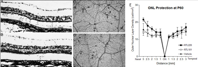
 Figure 1 of
Adamus, Mol Vis 2012; 18:2323-2337.
Figure 1 of
Adamus, Mol Vis 2012; 18:2323-2337.