Figure 2. Cdh23 transcripts
are expressed in the zebrafish brain and retina. In situ
hybridization showing cadherin 23 mRNA (mRNA) expression
in the developing and mature zebrafish nervous system. A:
Dorsal view of a zebrafish brain at 24 h postfertilization
(hpf); the blue/purple signal shows the first detectable
expression of cdh23 mRNA in two paired nuclei in the
diencephalon, adjacent to the eye. B: Ventral view of
intensely stained deep nuclei in the ventral diencephalon (vd)
at 48 hpf. C: Lateral view of 48 hpf larva. The two most
prominent nuclei labeled are located in the olfactory organ (oo)
and the ventral diencephalon. Additional brain labeling is also
observed, as is labeling of hair cells in the otic vesicle (ov).
D: Sense control shows absence of nonspecific labeling. E:
Dorsal view of the brain at 54 hpf, showing telencephalic and
ventral diencephalic paired nuclei from a different angle, as
well as additional dispersed cells in the mesencephalon and
rhombencephalon. F: In the eye, a very small subset of
inner layer cells is labeled. G-K: Post–in situ
hybridization cryosectioning allowed the more accurate
identification of labeled brain nuclei. Paired telencephalic
nuclei were located just medial to the olfactory bulb (ob), in
the subpallium (G). Caudal to the anterior commissure
(ac), very intense labeling was found in bilateral bar-shaped
structures in the eminentia thalami (EmT). H, I:
Continuing caudally (J), bar-shaped nuclei became more
globular in the ventral thalamus. Scattered labeling was also
observed in the dorsal thalamus and points caudal in J-K.
L: Blue lines represent planes of cryosections made in G-K.
M: Cdh23 in situ hybridization in 6 weeks
postfertilization (wpf) zebrafish shows ventral and dorsal
thalamic labeling similar to that seen in larval stages, but is
expanded to even more ventral areas, and to cells lying just
ventral to the optic tectum (TeO). N, O: At 6
wpf and 6 months postfertilization (mpf), retinal labeling is
still restricted to a very small subpopulation of amacrine
cells. gcl, ganglion cell layer; inl, inner nuclear layer; ipl,
inner plexiform layer; is, inner segment; onl, outer nuclear
layer; opl, outer plexiform layer; os, outer segment. Scale
bars=100 µm (G-K, M), 50 µm (N), 20
µm (O).
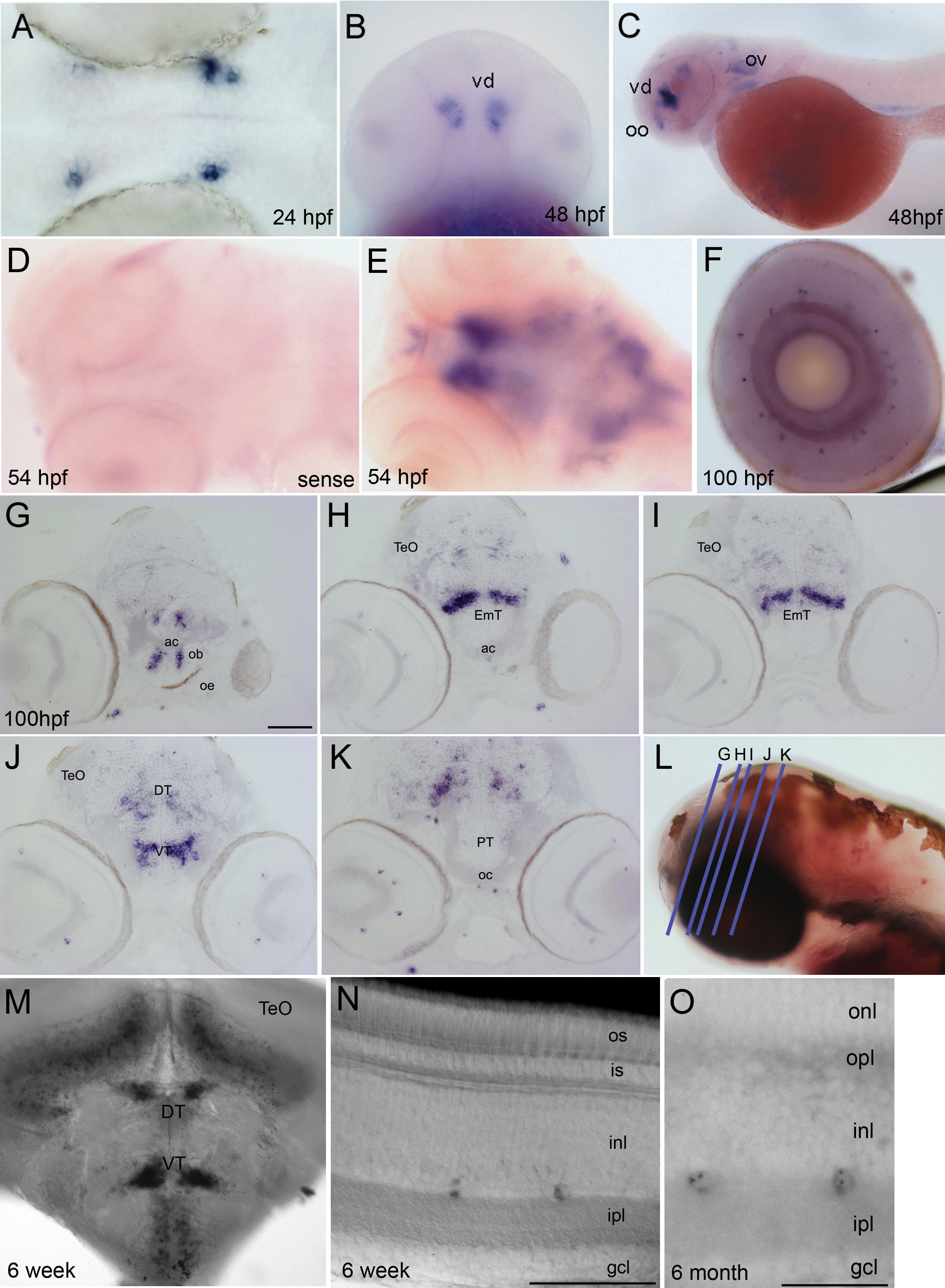
 Figure 2
of Glover, Mol Vis 2012; 18:2309-2322.
Figure 2
of Glover, Mol Vis 2012; 18:2309-2322.