Figure 1. Presence of full-length and
splice variants of cdh23 mRNA in zebrafish larval and
adult retina. A: Schematic illustrating the locations of
the mutations in the two cdh23 mutant fish lines,
reverse transcriptase (RT)–PCR primers, and the 5′ and 3′ in
situ hybridization probes used in this study. For the RT–PCR
primers, the red and orange arrows indicate the location of the
nested primer set used to amplify cdh23 in panels B
and C, while the cyan arrows indicate the primer set
used in D. B: Developmental time-course showing
the full-length cdh23 RT–PCR product isolated from eyes
of various ages (n=3). C: Smaller RT–PCR products were
also present. A shorter variant containing the transmembrane
(TM) segment was amplified from 7 days postfertilization (dpf)
and 6 months postfertilization (mpf) retinal transcripts (n=2).
A short, soluble variant was amplified from 6 weeks
postfertilization (wpf) eyes (n=2). Neither shorter variant was
isolated from 3 years postfertilization (ypf) eyes (n=2). D:
Using primers directed against sequence encoding the entire
C-terminus, both the full-length version (containing exon 68)
and the version present in the shorter variant containing the TM
segment were isolated from eye or enucleated head (n=2). E:
Schematics depicting the sequence and expected size of the
full-length and two short forms of cdh23 isolated in (B)
and (C).
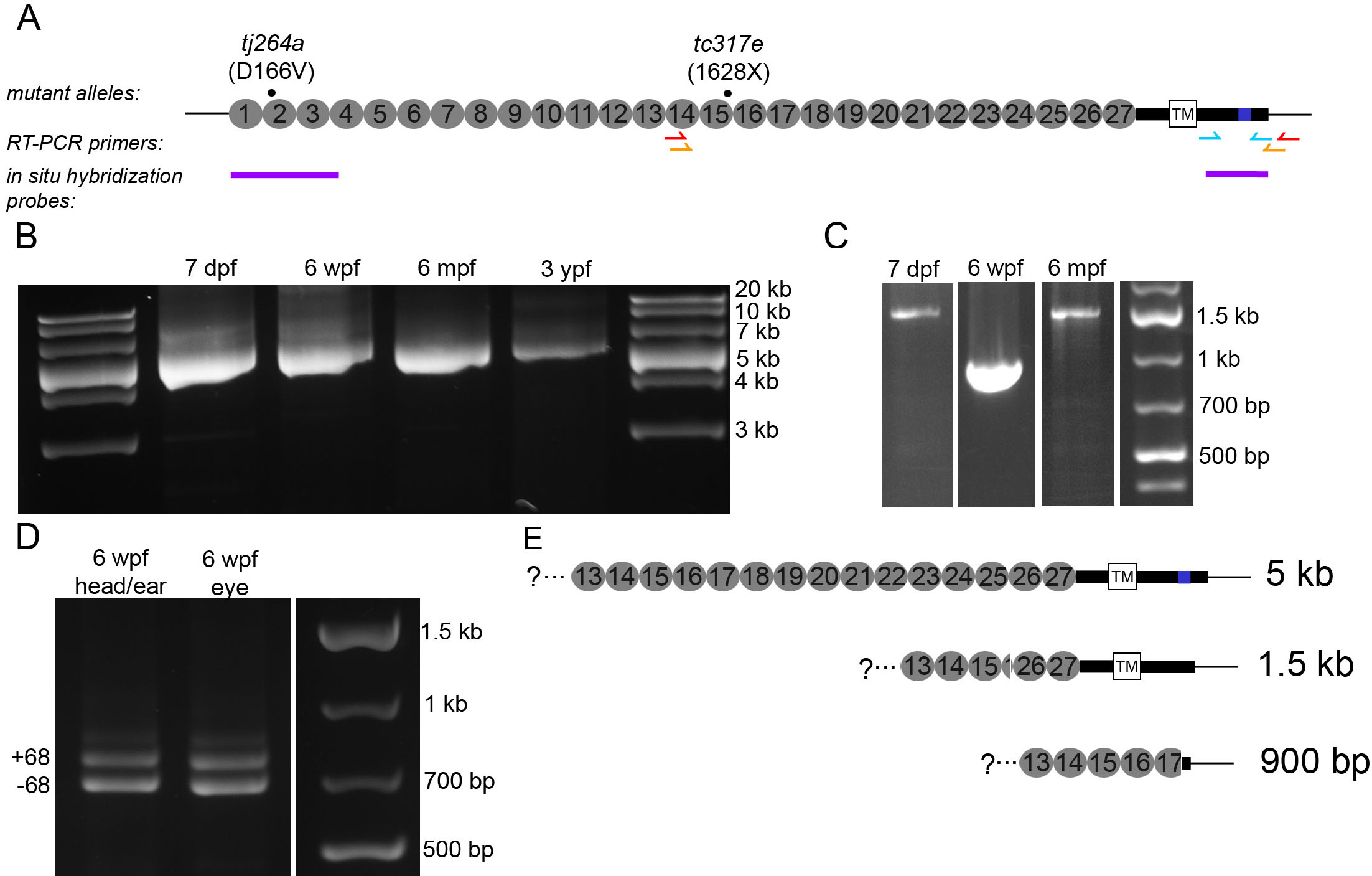
 Figure 1
of Glover, Mol Vis 2012; 18:2309-2322.
Figure 1
of Glover, Mol Vis 2012; 18:2309-2322.