Figure 3. Cytopathic effect of Acanthamoeba
on corneal fibroblasts in various conditions. A: Acanthamoebae
(0 to 10×103) were added to corneal fibroblasts in
each well and incubated at 25 °C for 2 days. Acanthamoebae
(1×10⁴) significantly decreased the viability of
corneal fibroblasts compared with no Acanthamoebae
(n=4). B: A significant decrease of corneal fibroblast
viability was detected from day 2 (n=4). C: Cytopathic
effect on corneal fibroblasts for 6 Acanthamoebae
isolates from our AK patients. A significant decrease of optical
density (indicating a cytopathic effect on corneal fibroblasts)
was detected with all tested Acanthamoebae compared to
control cultures with no Acanthamoebae (n=4). Similar
findings were obtained with repeated two experiments.
Representative data are shown. *p<0.05.
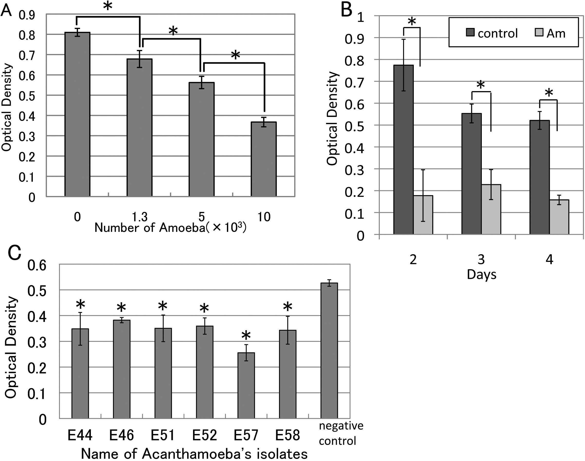
 Figure 3
of Takaoka-Sugihara, Mol Vis 2012;
18:2221-2228.
Figure 3
of Takaoka-Sugihara, Mol Vis 2012;
18:2221-2228.