Figure 2. Direct and indirect
cytopathic effects of Acanthamoeba on corneal
fibroblasts. A: Acanthamoebae were placed on
corneal fibroblasts at the center of 6-cm dishes and incubated
at 25 °C for 2 days. The fibroblasts are uniformly stained
with Giemsa solution in a control dish (left). The central area
where Acanthamoebae were placed shows no staining
(indicating loss of corneal fibroblasts) in a treated dish
(right). B: MTT assay showed there was no significant
difference of optical density value in the outer dishes with
corneal fibroblasts with or without insert culture dishes
bearing Acanthamoebae. Significant low optical density
value is detected in Acanthamoebae direct adhesion
group, compared with insert culture dishes bearing Acanthamoebae.
(n=6) Amoeba; Acanthamoeba, Inserter; insert culture dish. C:
Phase contrast microscopy shows many corneal fibroblasts are
detached and Acanthamoebae adhere to corneal fibroblasts
and the dish surface. Arrowheads show active Acanthamoebae
co-cultured with corneal fibroblasts. D: Confluent human
corneal fibroblasts are seen. Acanthamoebae in the
insert culture dishes with 0.4 µm pores are not observed.
Similar findings were obtained with repeated two sets of
experiments. Representative data are shown. Scale bar=10 µm.
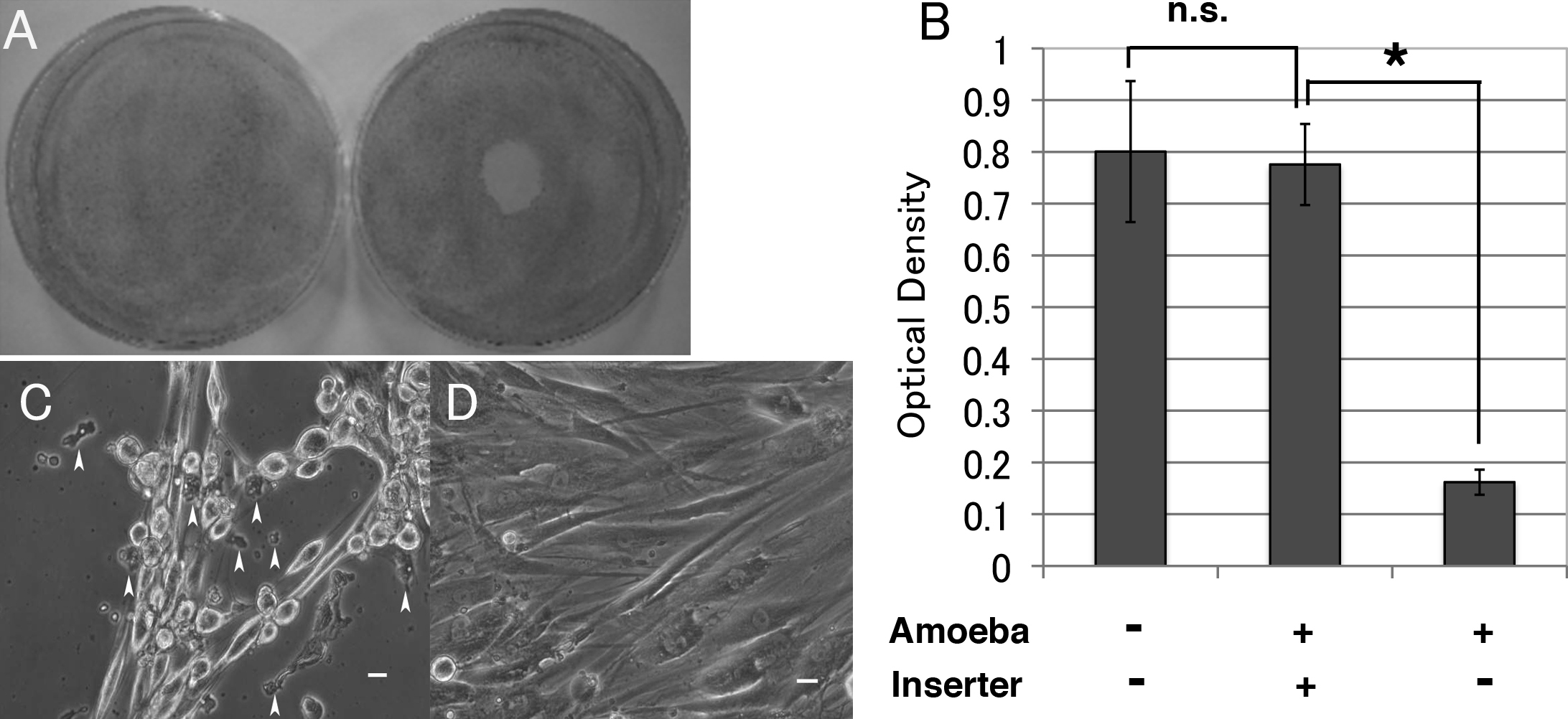
 Figure 2
of Takaoka-Sugihara, Mol Vis 2012;
18:2221-2228.
Figure 2
of Takaoka-Sugihara, Mol Vis 2012;
18:2221-2228.