Figure 1. Ex vivo invasion of Acanthamoeba into corneal stroma. Acanthamoebae were added to denuded human corneal stroma. Acanthamoebae were placed on the denuded corneal stroma (endothelial side up) and incubated at 25 °C for 2 days. Hematoxylin and eosin
staining shows Acanthamoebae (arrowheads) located in fine collagen fibrils. Arrow shows the direction of corneal epithelium. Scale bar=10 µm.
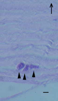
 Figure 1 of
Takaoka-Sugihara, Mol Vis 2012; 18:2221-2228.
Figure 1 of
Takaoka-Sugihara, Mol Vis 2012; 18:2221-2228.