Figure 1. HCE wound healing evaluation from Day 1 to Day 3. Effects of PBS (control; column 1), CsA-CEm (column 2), CsA-AEm (column
3), CsA-Oil (column 4), and 0.02% BAK solution (column 5) on HCE cell wound closure at D0, D1, D2, and D3. Closure of the
wound is observed for PBS and the three CsA groups at D1 and progresses until D3. 0.02% BAK treatment did not result in observable
wound closure; on the contrary, it induced cell death and widening of the wound. The solid lines indicate the initial WD,
and the dashed lines indicate the new edge positions at the different time points.
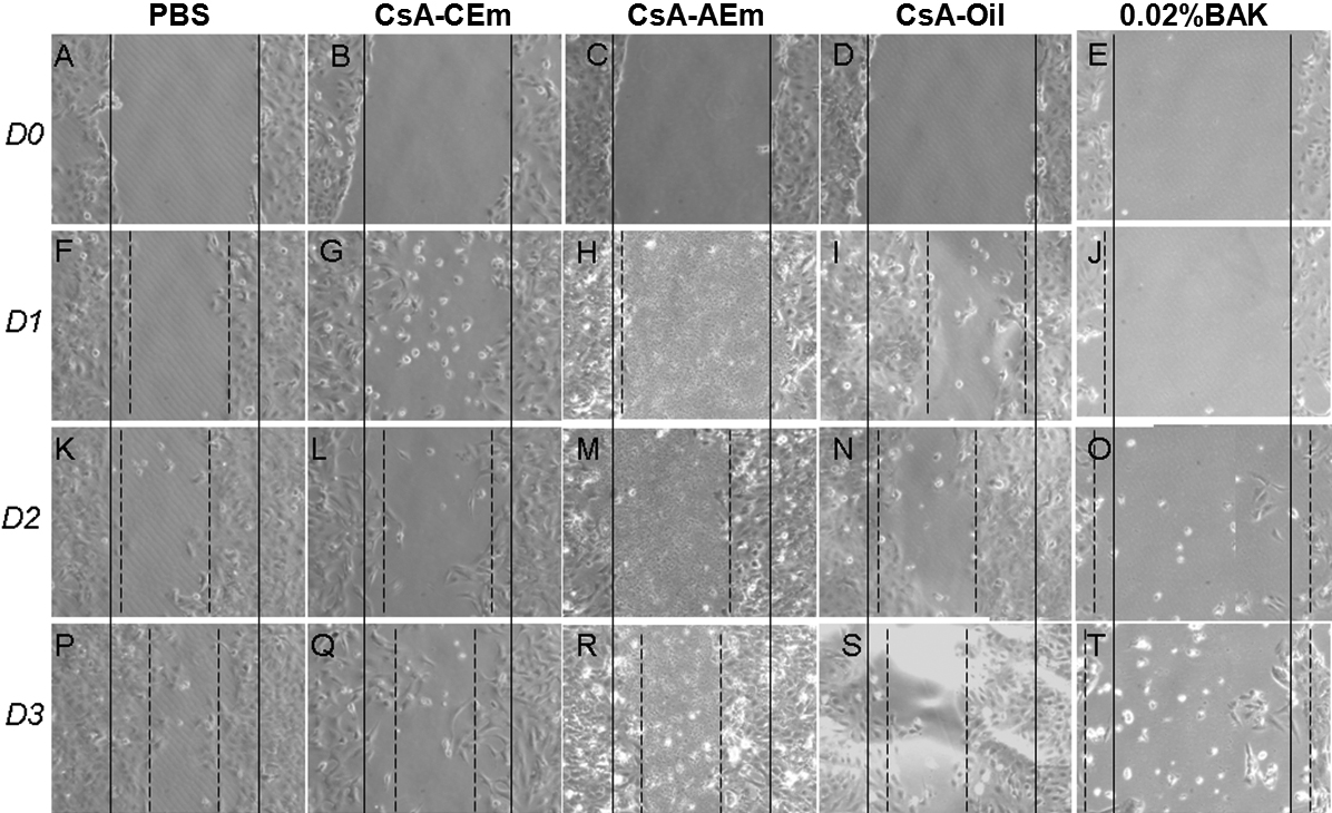
 Figure 1 of
Liang, Mol Vis 2012; 18:2195-2204.
Figure 1 of
Liang, Mol Vis 2012; 18:2195-2204.