Figure 1. Anterior segment photographs of case 1 and case 2 patients with aniridia are shown. A: Shows the case 1 patient (25 years old, male), who had very serious cataracts and corneal degeneration, in addition to aniridia
of the right eye. B: Shows the left eye of patient 1, who had partial cataracts. C and D: Show anterior segment photographs of both eyes of case 2. Some pigment particles precipitated in the front of the anterior
lens capsule.
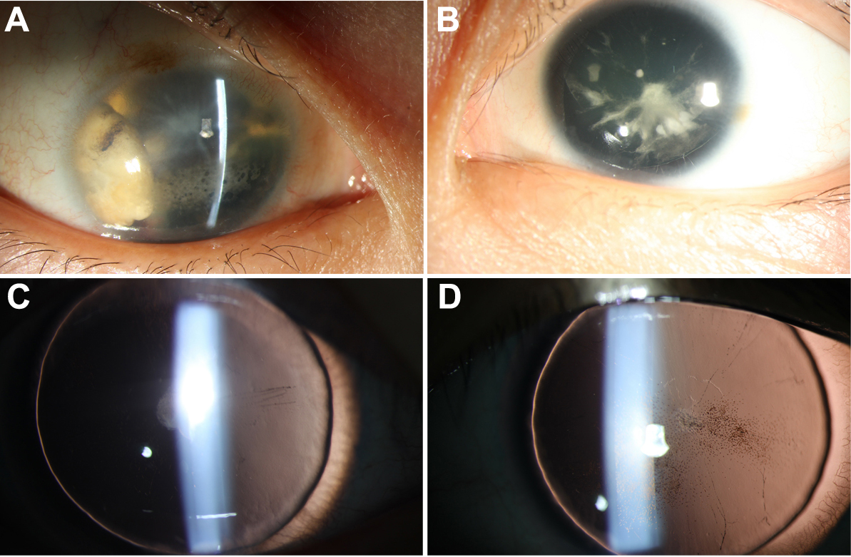
 Figure 1 of
Lin, Mol Vis 2012; 18:2190-2194.
Figure 1 of
Lin, Mol Vis 2012; 18:2190-2194.