Figure 5. Imaging of the
morphological dynamics of posterior fiber cells following UVA
irradiation. The morphological alterations of the fibers after 5
min (A), 30 min (B), 3 h (C), and 18 h (D)
are shown. The round irradiation area (ring) exhibited no
obvious changes after 5 min. The fibers on this area aggregated
into globules after 30 min (B, C). After 18 h,
the aggregation area enlarged to a triquetrous opaque and showed
a liquid tremor (D). Three plaques of damage appeared on
the adjacent area after 5 min (A), which became more
severe after 30 min (B). This damage gradually faded, and
it completely disappeared after 18 h (C, D).
High-magnification images of the fiber changes after 30 min are
shown in E and F. There are two different types
of damages: the fibers on the irradiation area aggregated into
globules (E, rectangle); the extracellular space between
the fibers on the adjacent area became enlarged, but the fibers
themselves did not break down (F, rectangle). Scale bars:
A–F=50 μm.
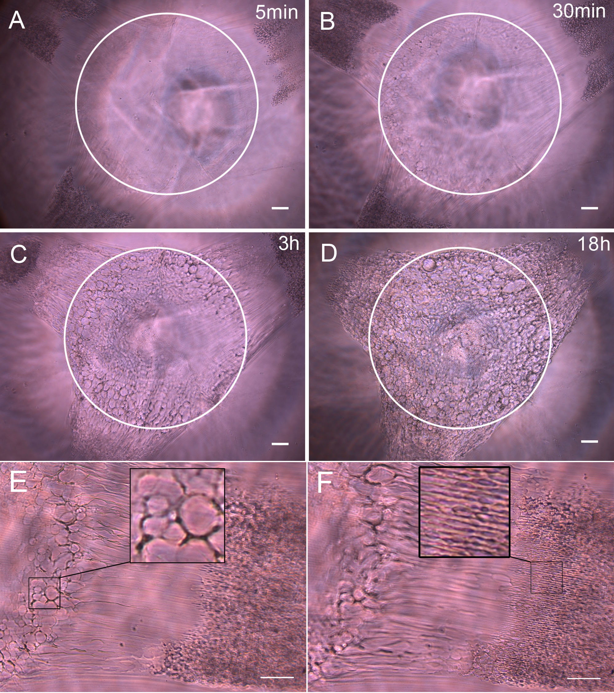
 Figure 5
of Kong, Mol Vis 2012; 18:2165-2173.
Figure 5
of Kong, Mol Vis 2012; 18:2165-2173.