Figure 3. Imaging of mitosis of the
LECs. Numerous mitotic figures were present all over the
anterior surface after 15 h of incubation with 20 ng/ml IGF-I (A).
The chromosomes and the profiles of the cell bodies were
distinctly visible, and dividing cells in various stages could
easily be observed (B–J). A vague, dark ring (arc)
was located in the center of the lens surface (A). The
curved surface of the lens permitted visualization of a small
area of cells in the focal plane. Scale bars A=50 μm; B–J=10
μm.
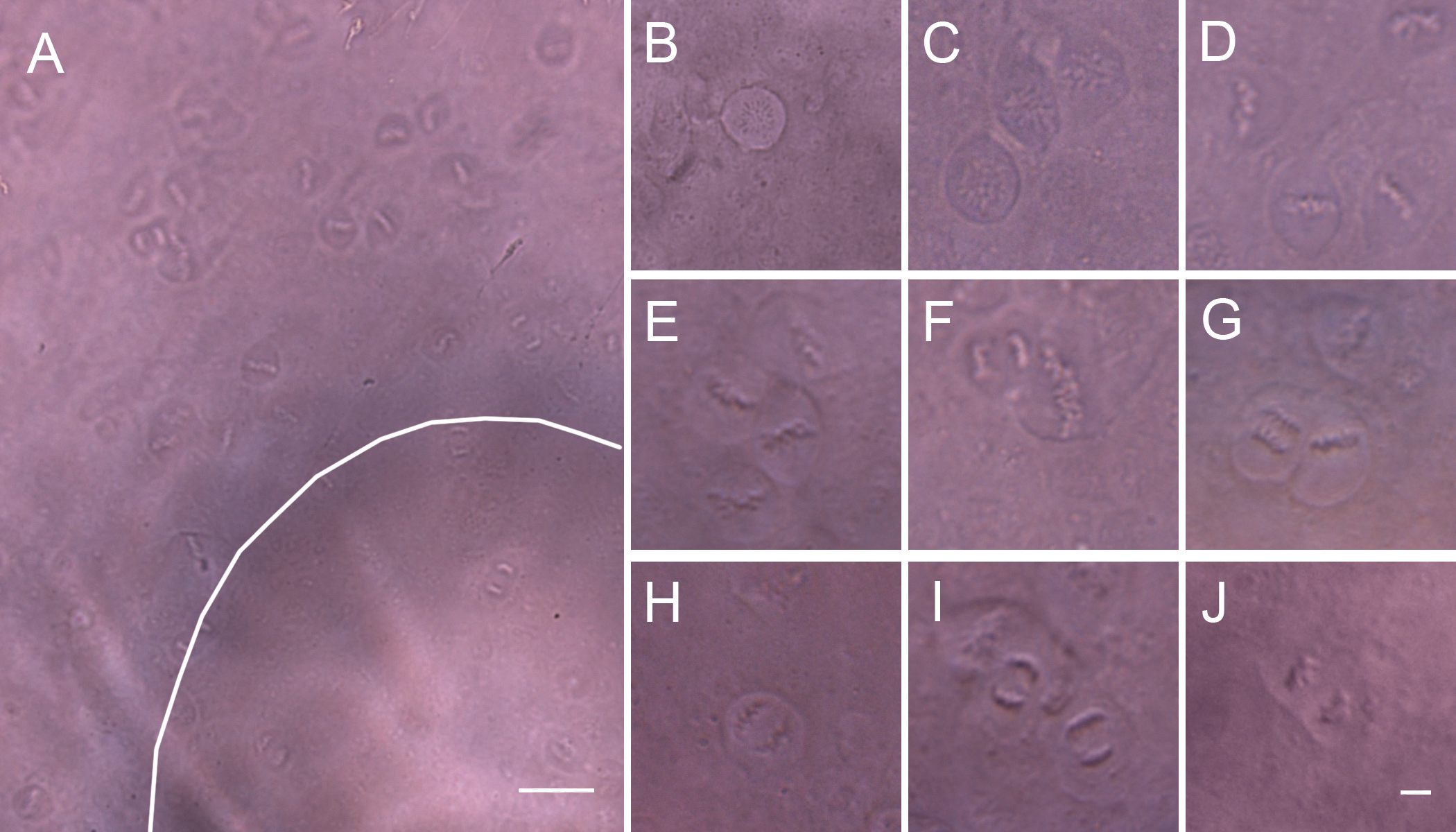
 Figure 3
of Kong, Mol Vis 2012; 18:2165-2173.
Figure 3
of Kong, Mol Vis 2012; 18:2165-2173.