Figure 2. Images of the anterior and
posterior surfaces of the lens. Small, rounded humps (arrow)
were present all over the anterior surface, which was the major
morphological feature of the LECs using our method (A).
The chromatin of the LECs was stained with Hoechst 33342 (B).
There were no humps on the posterior surface of the lens. The
Y-suture (arrow) and the superficial fiber cells were clearly
revealed (C). After two hours out of the incubator, the
lens epithelial layer exhibited some regional changes near the
equator (D, ring). Scale bars: A-C=10 μm;
D=50 μm.
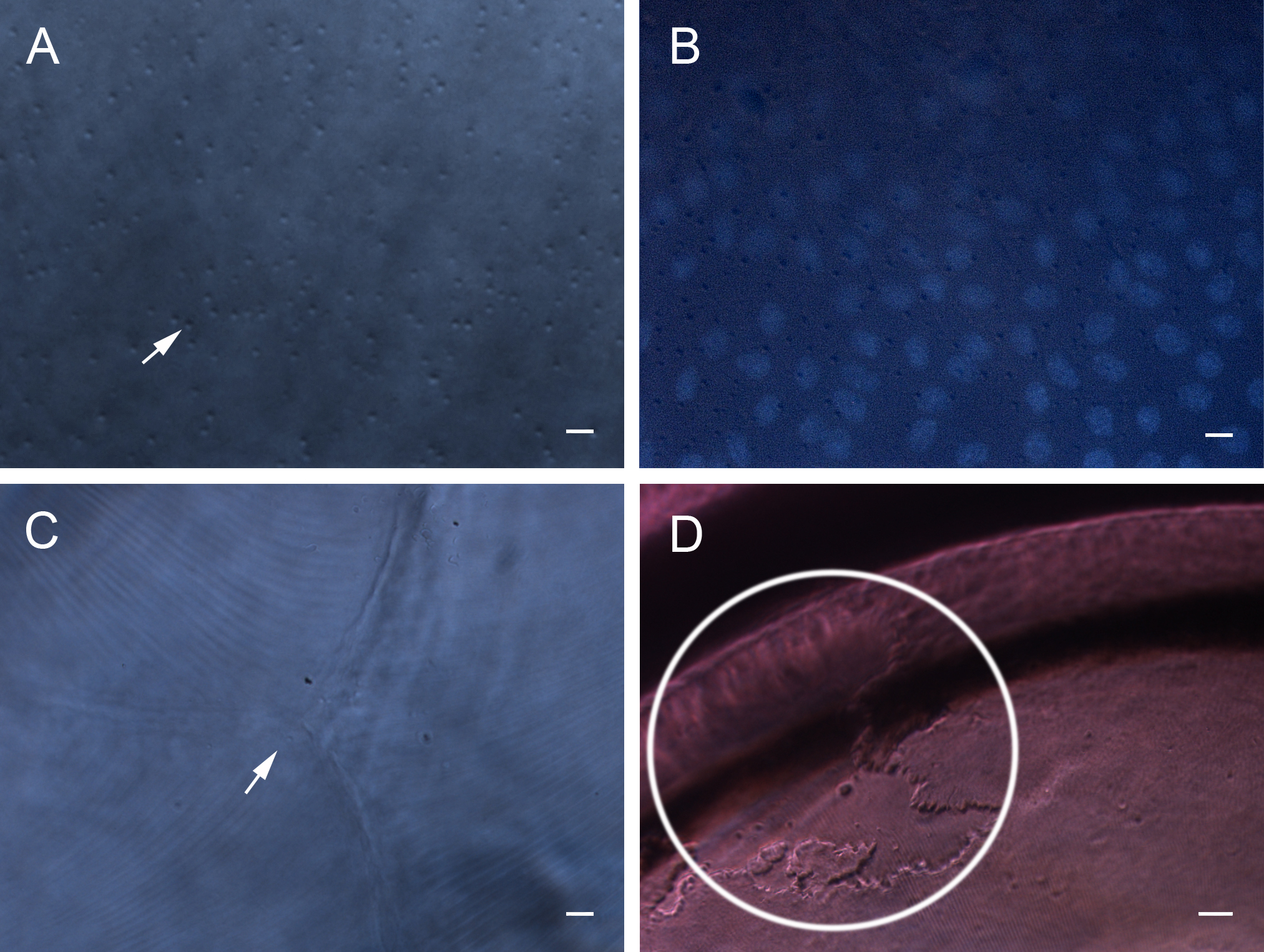
 Figure 2
of Kong, Mol Vis 2012; 18:2165-2173.
Figure 2
of Kong, Mol Vis 2012; 18:2165-2173.