Figure 4. Following PMA treatment, HuR protein translocates from the nuclear area to the cytoplasm in pericytes and endothelial cells.
Confocal fluorescence representative images of HuR in control (CTR) and PMA-treated (PMA) retinal pericytes (PC) and endothelial
cells (EC) in coculture. The arrows indicate that HuR protein is localized mainly in the nuclear area in untreated cells,
and migrates to the cytoplasm following PMA stimulus. Scale bars: 20 μm.
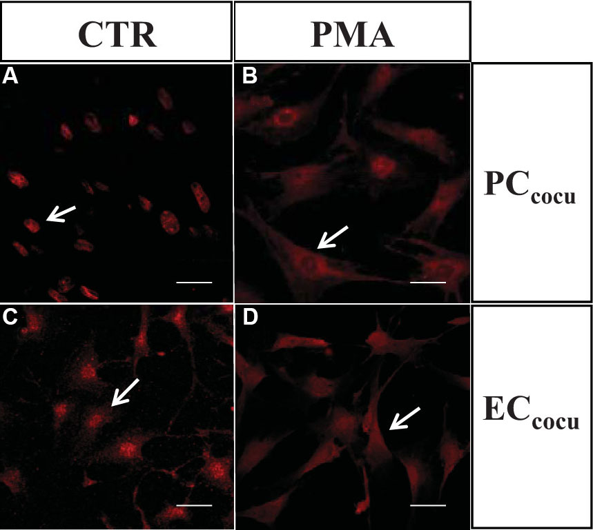
 Figure 4 of
Amadio, Mol Vis 2012; 18:2153-2164.
Figure 4 of
Amadio, Mol Vis 2012; 18:2153-2164.