Figure 2. VEGF protein levels are
affected by culture conditions in pericytes and endothelial
cells. A: Representative real-time reverse
transcriptase–PCR amplification plots relative to VEGF
mRNA content in pericytes (PC) and endothelial cells (EC; Ct
mean±SEM: PC: 26.9±1.1; EC: 28.8±1.7). B: Representative
western blotting of VEGF protein in the total homogenates of
pericytes and endothelial cells cultured separately (PCmono
and ECmono, respectively). C-D: Mean
gray levels ratios (mean±SEM) of VEGF/α-tubulin
immunoreactivities measured by western blotting in PC (C)
and EC (D). All the comparisons were performed between
cells in monoculture (mono) and cells in coculture (cocu).
*p<0.05; **p<0.01, n=4. E: VEGF protein levels
measured in cell culture conditioned media of PCmono,
ECmono, and coculture. **p<0.01; ***p<0.001,
n=4, Tukey-Kramer test.
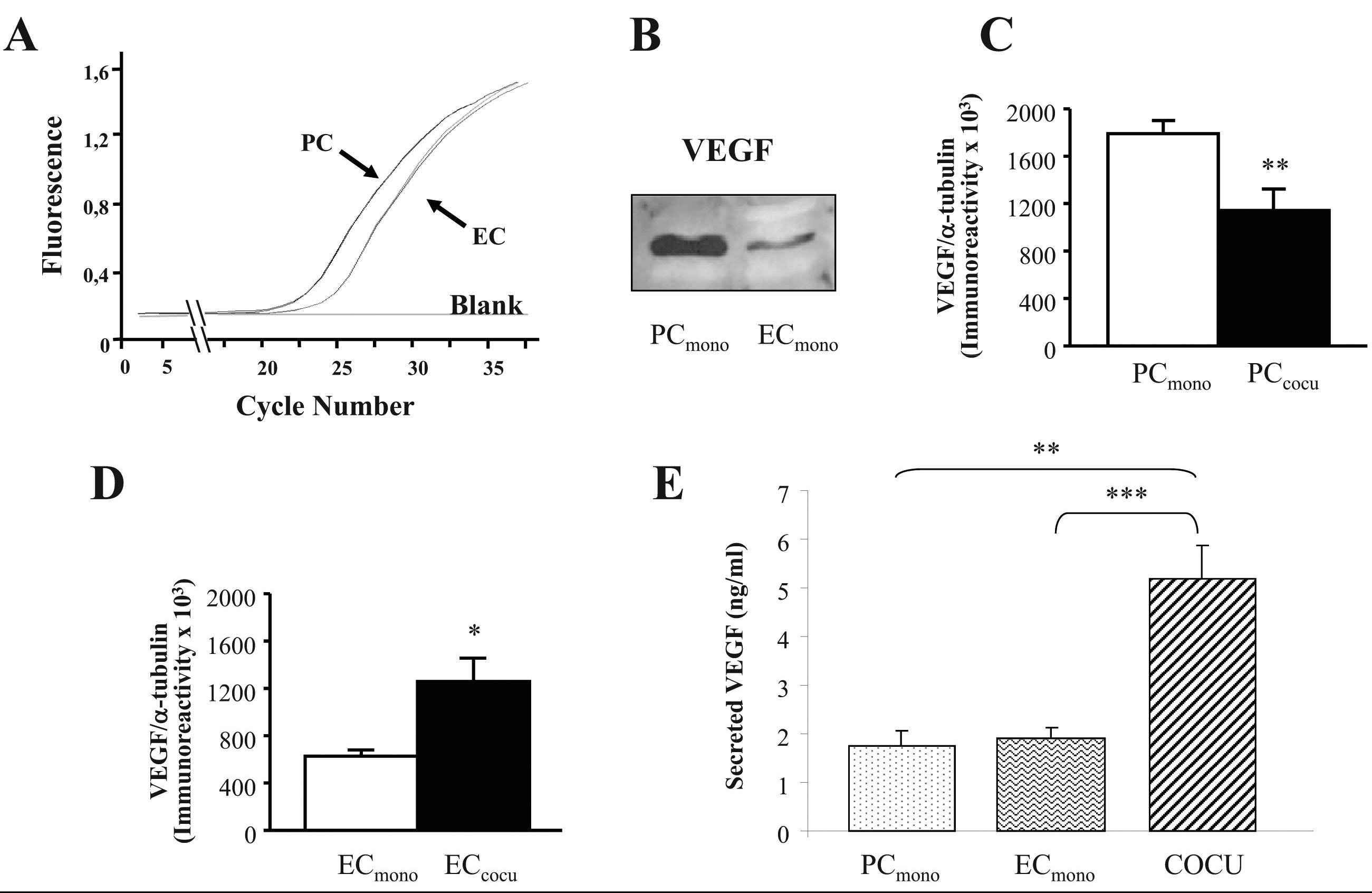
 Figure 2
of Amadio, Mol Vis 2012; 18:2153-2164.
Figure 2
of Amadio, Mol Vis 2012; 18:2153-2164.