Figure 2. Optical coherence
tomography Doppler imaging before and after optic nerve crush
(ONC). OCT was used to measure blood flow using Doppler imaging.
We were able to confirm that there is continued perfusion of the
retina before and after ONC. Panel A shows the OCT
Doppler image of the retina immediately before ONC, and panel B
illustrates the blood flow immediately after ONC. The arrows
point to some of the vessels present on the surface of the
retina. The colors reflect the direction of blood flow; red is
flow toward the transducer and blue is flow away from the
transducer. These data demonstrate that in the mouse this
procedure does not interrupt blood flow to the retina. The
abbreviations in the figures are defined as follows: retinal
pigment epithelium (RPE), outer nuclear layer (ONL), inner
nuclear layer (INL), and ganglion cells (GC).
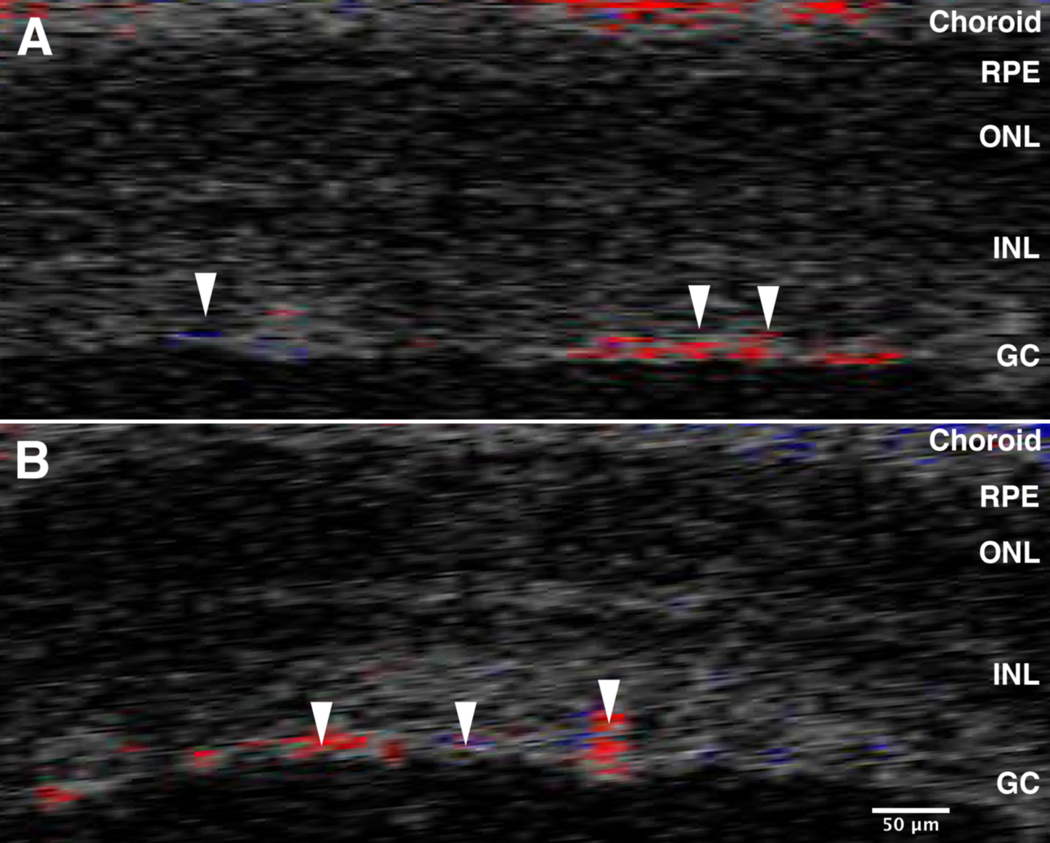
 Figure 2
of Templeton, Mol Vis 2012; 18:2147-2152.
Figure 2
of Templeton, Mol Vis 2012; 18:2147-2152.