Figure 1 of
Batista, Mol Vis 2012; 18:194-202.
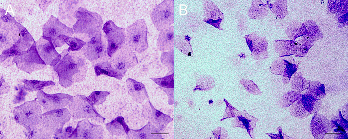
Figure 1.
Impression cytology of the corneas of control (
A
) and aged (
B
) rats. Grades from 0 to 3 were given for each sample, based on size, nucleus, and the presence of mucus (Scale bar=25 µm).
 Figure 1 of
Batista, Mol Vis 2012; 18:194-202.
Figure 1 of
Batista, Mol Vis 2012; 18:194-202.