Figure 2. Clonogenic properties and proliferative potential of L-MSC. The cells were cultured for 10 days and stained with 0.5% crystal
violet in methanol. A colony-forming unit was determined when measured >2 mm (arrow). Colonies that measured <2 mm or faintly
stained were excluded (A). B: Representative growth curve of L-MSC as a function of time after the culture. Data are expressed as mean±SEM from three
independent experiments.
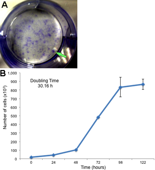
 Figure 2 of
Garfias, Mol Vis 2012; 18:2087-2095.
Figure 2 of
Garfias, Mol Vis 2012; 18:2087-2095.