Figure 1. Immunophenotypical characterization of L-MSC. Cells at the 3rd passage were trypsinized, labeled with antibodies against the
indicated antigens and analyzed by flow cytometry. As shown, the cells weakly expressed vimentin, meanwhile, they were positive
to CD29, CD34, CD39, CD73, and CD105. On the other hand, the cells were negative to cytokeratin, HLA-DR, and CD45 expression.
Representative dot plots from 3 separate samples are shown.
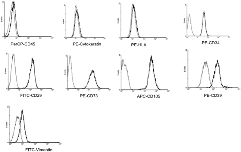
 Figure 1 of
Garfias, Mol Vis 2012; 18:2087-2095.
Figure 1 of
Garfias, Mol Vis 2012; 18:2087-2095.