Figure 2. Analysis of LEP503 promoter
specificity. A: The expression of EGFP driven by lenti-LEP503-HSV-tk-EGFP
vector in HLECs, RPECs, and ASFCs at 72 h after being infected.
B: The expression of EGFP driven by lenti-CMV-HSV-tk-EGFP
vector in HLECs, RPECs, and ASFCs at 72 h after being infected.
Magnification: 40×; Green: EGFP, Blue: DAPI staining. C:
Flow cytometry evaluation of the expression of EGFP driven by
lenti-LEP503-HSV-tk-EGFP and lenti-CMV-HSV-tk-EGFP
vectors in HLECs, RPECs, and ASFCs at 48 h after being infected.
D: western blotting assessment of the expression of EGFP
driven by lenti-LEP503-HSV-tk-EGFP vector at 24, 48, and
72 h after being infected, and by lenti-CMV-HSV-tk-EGFP
vector 72 h after being infected in HLECs, RPECs, and ASFCs. E:
Semi-quantitative analysis of the western blotting results. The
intensities of HSV-tk mRNA were normalized to ACTB
(n=3).
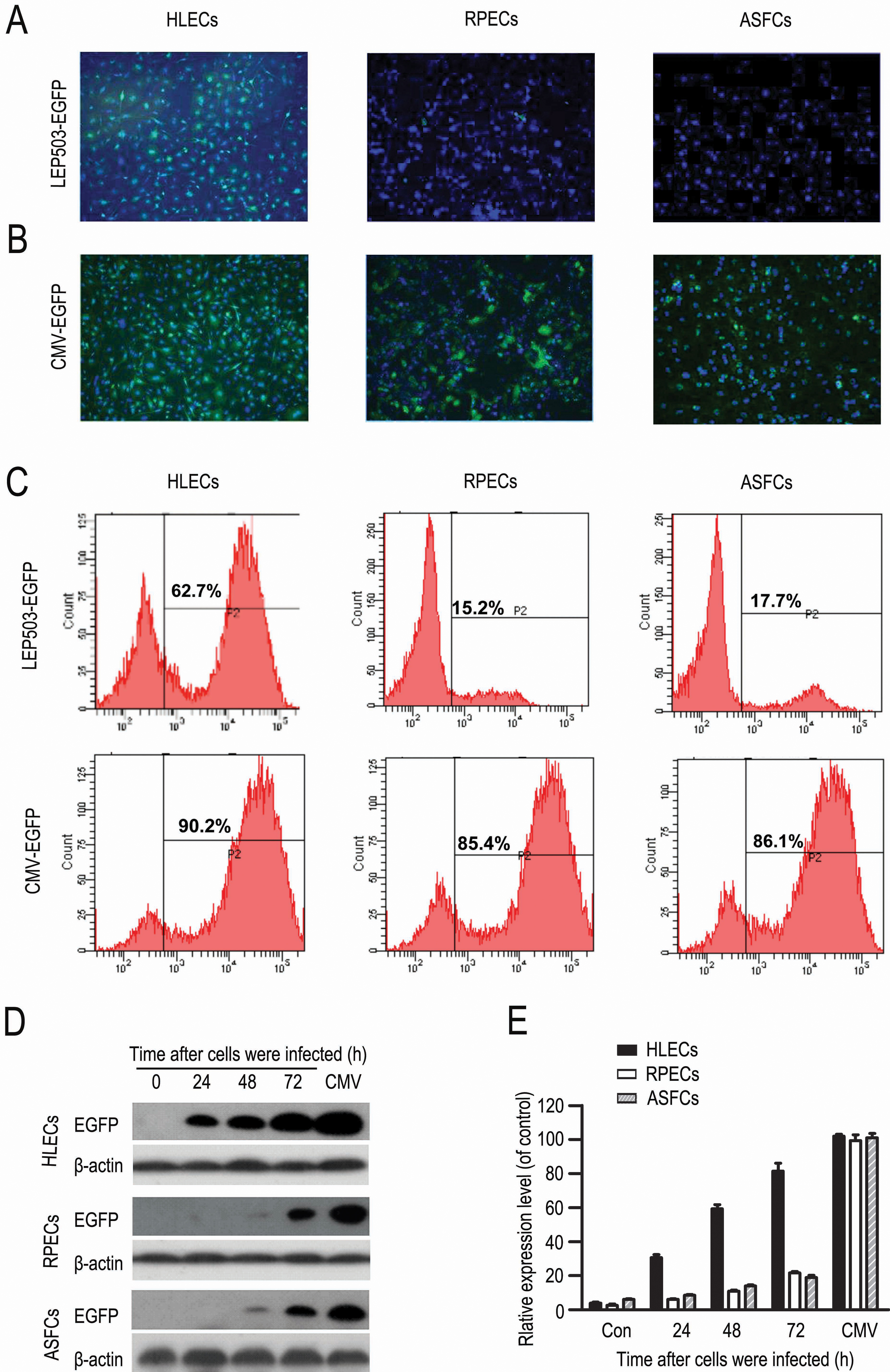
 Figure 2
of Yang, Mol Vis 2012; 18:2053-2066.
Figure 2
of Yang, Mol Vis 2012; 18:2053-2066.