Figure 2. Clinical features of the affected individuals. A: Photograph of affected individual IV-3. Note the classical Gower’s sign on trying to get up from the sitting position. B: Fundus photograph of affected individual IV-3. Note the depigmented retina and underlying choroid vessels. C-F: Transverse sections of skeletal muscle: C and D from an unrelated normal individual and E and F from affected individual IV-3. Note normal polygonal myofibers with peripheral nuclei and uniform diameter in panel C (hematoxylin and eosin staining) and normal positive immunostaining of DMD protein along the sarcolemma in all the fibers
in panel D. Note rounding, variation in diameter, central nuclei, regenerating fibers and fibrosis in panel E (hematoxylin and eosin staining), and total absence of DMD staining in all the fibers in panel F. G: Partial facial photograph of affected individual IV-3 showing blue iris. H: Partial facial photograph of affected individual IV-2 showing heterochromia of iris. I: Partial facial photograph of paternal grandmother II-2 showing heterochromia of iris.
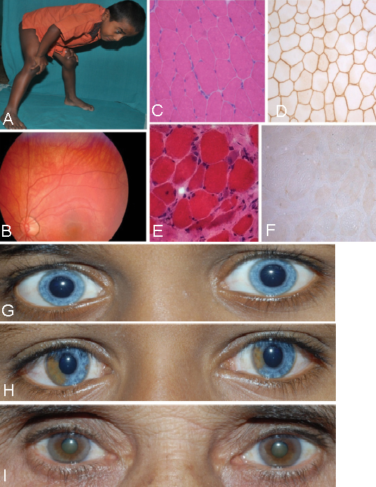
 Figure 2 of
Kapoor, Mol Vis 2012; 18:2022-2032.
Figure 2 of
Kapoor, Mol Vis 2012; 18:2022-2032.