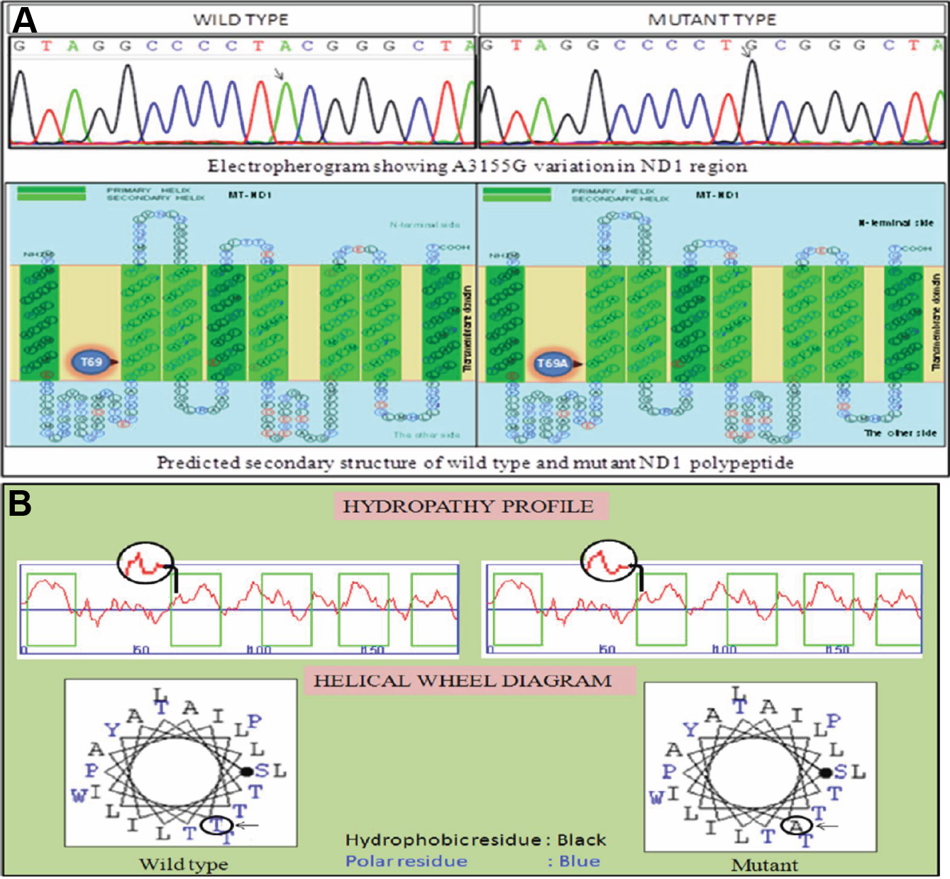
Figure 5. Electropherogram and polypeptide of wild type and mutant sequence of ND1 region.
A: Electropherogram showing wild type and mutant nucleotide sequence of ND1 region with A>G transition at 3155 nucleotide position.
Predicted secondary structure of the wild type and mutant ND1 polypeptide showing the altered amino acid.
B: The architecture of the membrane proteins is reflected in the hydropathy profile. The helical wheel diagram of predicted
polypeptide show change from polar residue to hydrophobic residue. Arrow mark indicates the altered aminoacid position. The
source of the structure prediction is
SOOSUI.
 Figure 5 of
Roshan, Mol Vis 2012; 18:181-193.
Figure 5 of
Roshan, Mol Vis 2012; 18:181-193.