Figure 5. Comparison of the expression patterns of matrilin-4 in healthy, granular type I, and lattice type I stromal dystrophy corneal
buttons. Some immunolabeling was recorded in healthy corneal epithelium (A). In granular dystrophy, all epithelial layers were positive for matrilin-4, and no Bowman’s layer was present under the
epithelial basement membrane (B). Altered epithelial cell morphology in the basal layer was observed, with strong basal immunolabeling for matrilin-4 in
lattice type I dystrophy (C). Scale bar 100 μm.
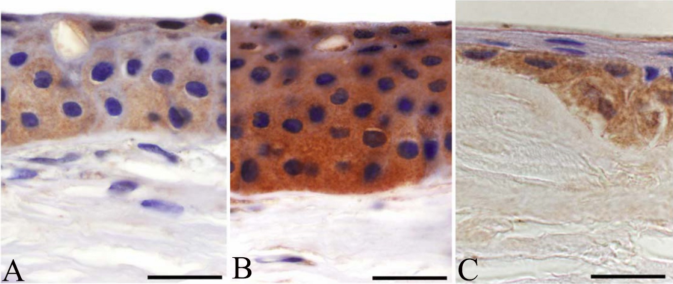
 Figure 5 of
Szalai, Mol Vis 2012; 18:1927-1936.
Figure 5 of
Szalai, Mol Vis 2012; 18:1927-1936.