Figure 4. Comparison of the expression patterns of matrilin-2 in healthy, granular type I, and lattice type I stromal dystrophy corneal
buttons. In normal controls, matrilin-2 was present in most corneal epithelial layers (A). Intense epithelial immunopositivity was observed in granular dystrophy. Focal thinning or loss of Bowman’s layer was also
apparent (B). Enlarged immunopositive basal cells in corneal epithelium with mild stromal staining were observed in lattice dystrophy
(C). Scale bar 100 μm.
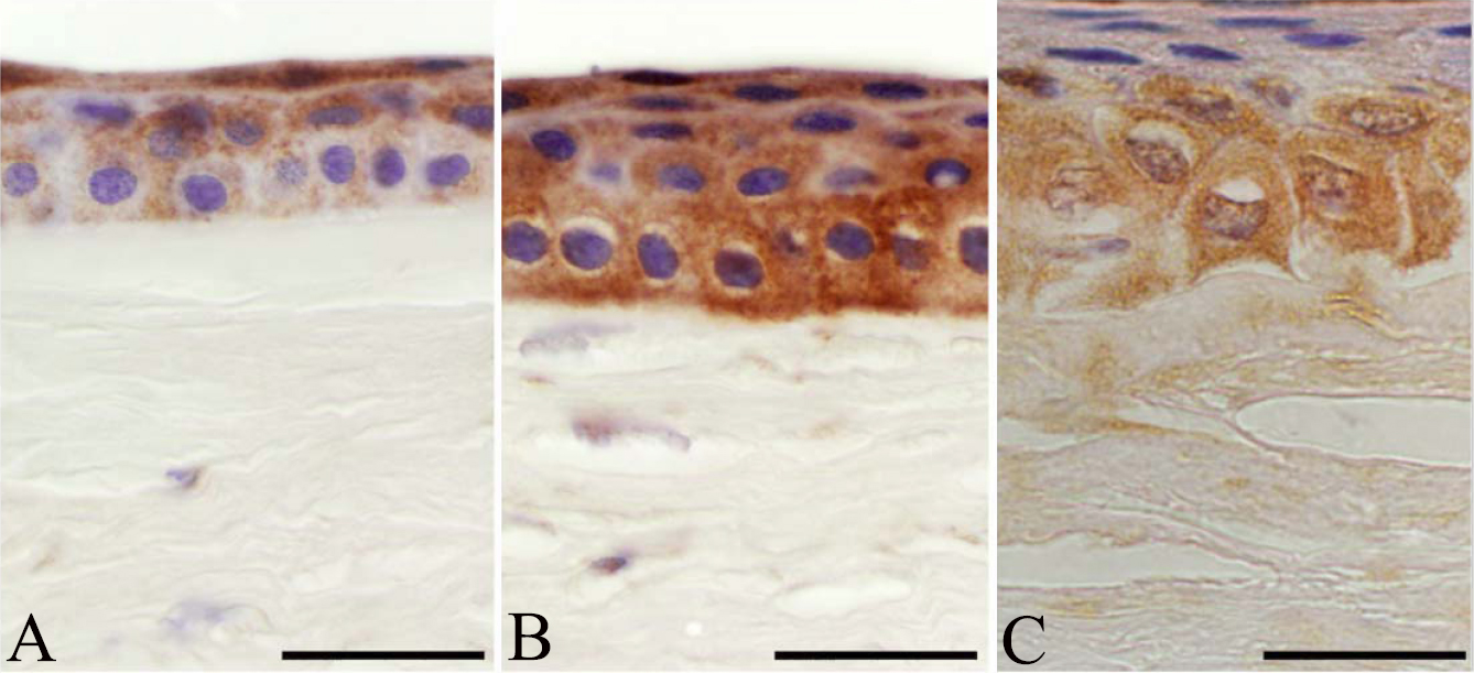
 Figure 4 of
Szalai, Mol Vis 2012; 18:1927-1936.
Figure 4 of
Szalai, Mol Vis 2012; 18:1927-1936.