Figure 3. Comparison of the expression patterns of tenascin-C in healthy, granular type I, and lattice type I stromal dystrophy corneal
buttons. Cytoplasmic immunolabeling in normal sections was only observed in some basal epithelial cells (A). In granular dystrophy, diffuse, marked immunostaining was recorded in the epithelial layers and in the stromal granules
(B). Basal epithelium showed moderate immunostaining for tenascin-C in lattice dystrophy (C). Scale bar 100 μm.
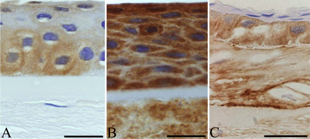
 Figure 3 of
Szalai, Mol Vis 2012; 18:1927-1936.
Figure 3 of
Szalai, Mol Vis 2012; 18:1927-1936.