Figure 2. Comparison of expression patterns of fibrillin-2 in healthy, granular type I and lattice type I stromal dystrophy corneal
buttons, and the colocalization of matrilin-2 and fibrillin-2 in corneal epithelium. Healthy samples showed mild immunolocalization
in superficial and basal epithelial layers (A). In addition to the strong epithelial immunopositivity of fibrillin-2 in granular dystrophy, marked labeling was observed
in stromal granules (B). In lattice corneal dystrophy, continuous cytoplasmic staining was found in the base of the basal columnar cells (C). Scale bar 100 μm.
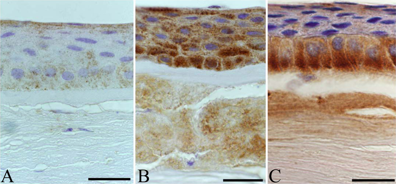
 Figure 2 of
Szalai, Mol Vis 2012; 18:1927-1936.
Figure 2 of
Szalai, Mol Vis 2012; 18:1927-1936.