Figure 1. Granular and lattice type I dystrophies: biomicroscopic and histopathologic findings. The clinical appearance of lattice corneal
dystrophy (A). The branching dots and lines, representing amyloid deposits, are typical in the corneal stroma. Granular corneal dystrophy
under slit lamp and retroillumination (B). Well defined, white granules are present in the central corneal stroma (C). Histological section of the granular type I corneal button (H&E) (D). The deposits are observed in the sub-Bowman layer in the stroma. The section of lattice corneal dystrophy stains weak with
Congo Red (E). Stromal fusiform amyloids stain with Congo red (F) and show birefringence under polarized light. The colocalization of matrilin-2 and fibrillin-2 in normal corneal epithelium
is presented in G-J. High magnification microphotos of single laser scanning confocal optical sections (1 μm) demonstrating the immunolocalization
of the anti-matrilin-2 (H, green) and anti-fibrillin-2 (I, red) antibody in corneal epithelial layer. The nuclei are blue on each images presented (G-J). Note that no nuclear staining is present on the merged image (J) preclude the possibility of nonspecific reaction (internal control). Around the smaller DAPI labeled nucleus yellow color
may indicate the colocalization of these two antibodies used. Scale bars for light and polarized microscopic images are 100
μm, for confocal images; 10 μm.
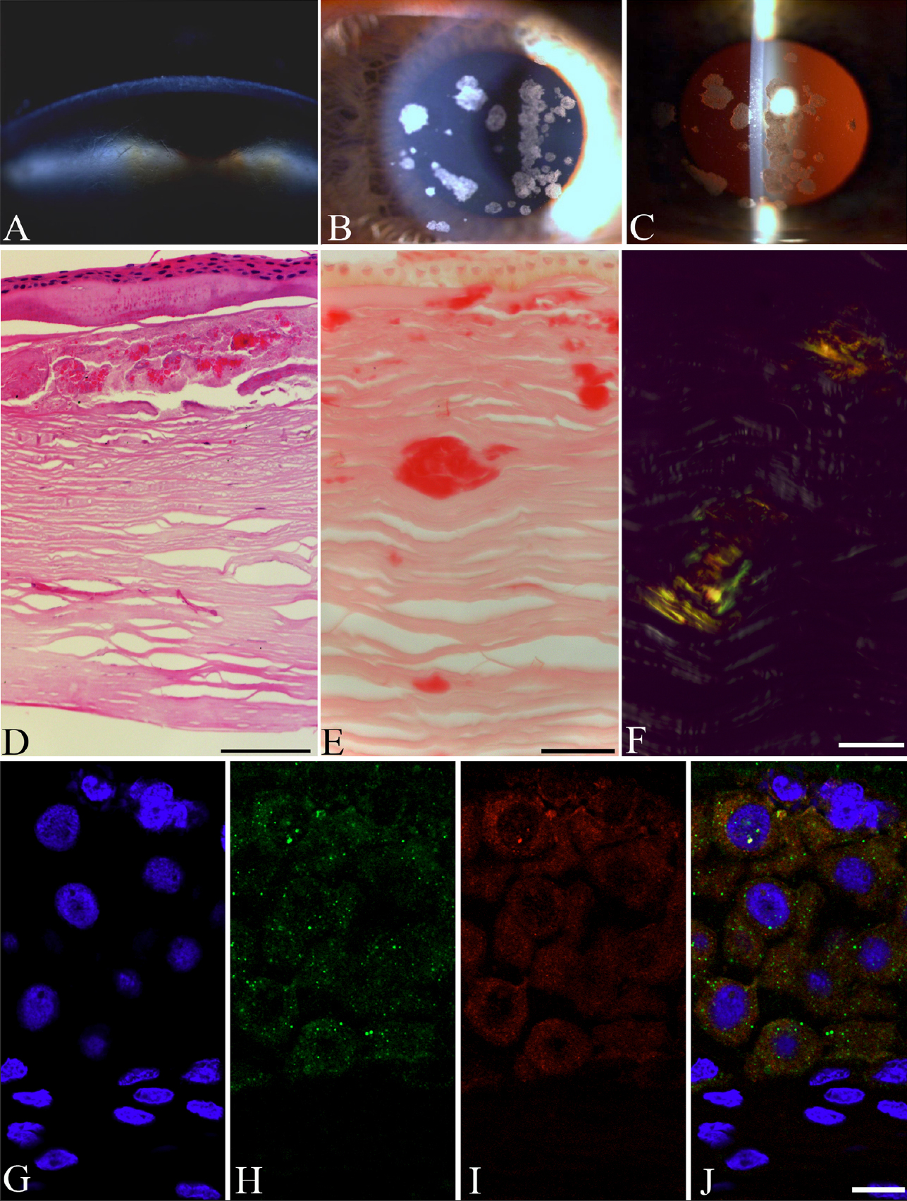
 Figure 1 of
Szalai, Mol Vis 2012; 18:1927-1936.
Figure 1 of
Szalai, Mol Vis 2012; 18:1927-1936.