Figure 6. TUNEL staining of retinal
EC. Retinal ECs were transfected with the pCMV plasmid
containing human caspase-14 cDNA and the empty pCMV vector as a
control (A). The number of apoptotic cells (green) was
significantly increased in caspase-14 expressing pericytes
compared to the control (B, C). DAPI (blue) is a
nuclear staining (*p<0.05).
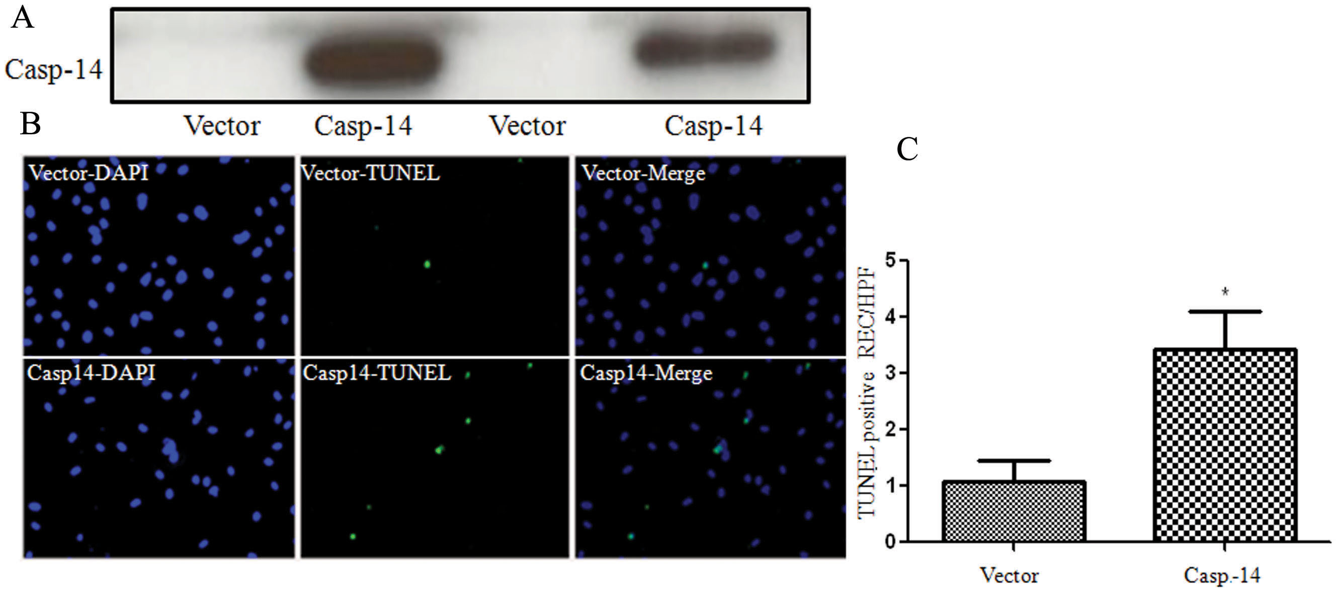
 Figure 6
of Al-Shabrawey, Mol Vis 2012; 18:1895-1906.
Figure 6
of Al-Shabrawey, Mol Vis 2012; 18:1895-1906.