Figure 3. Caspase-14 in the mouse
retina. Immunofluorescence reaction (A) using specific
antibody against caspase-14 (green), vascular marker (isolectin
B4-red), and nuclear stain (DAPI) showed marked increase in the
expression of caspase-14 in different layers in particular in
relation to retinal vasculatures (red). Western blot analysis of
caspase-14 in the mouse retina (B) demonstrated
significant upregulation in diabetic mice and the presence of
cleaved caspase-14 in one of the diabetic mice compared to the
control (C). (* p<0.05; D versus C; n=6).
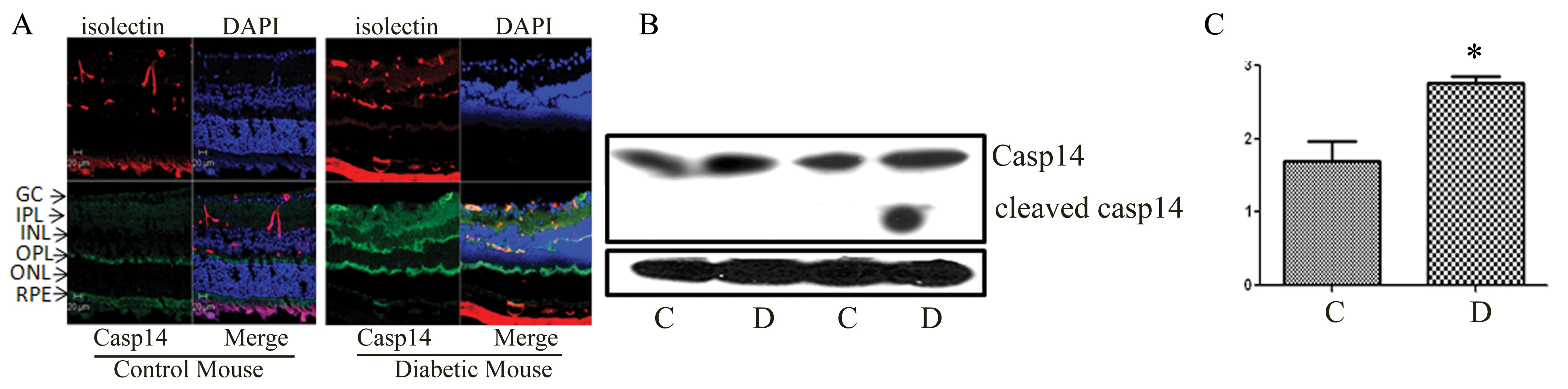
 Figure 3
of Al-Shabrawey, Mol Vis 2012; 18:1895-1906.
Figure 3
of Al-Shabrawey, Mol Vis 2012; 18:1895-1906.