Figure 3. Novel retinal findings. Panels A and B are of fundus photos showing a normal-looking macula and an abnormally mottled retinal pigment epithelium in the periphery.
The photo in Panel C shows the demarcation line circular, separating the normal-looking macula from abnormal periphery, mild optic nerve atrophy;
optic nerve pit and attenuation of retinal arterioles could be seen (Patient 3). Panel D shows the proband’s local macroaneurysm with limited retinal hemorrhage.
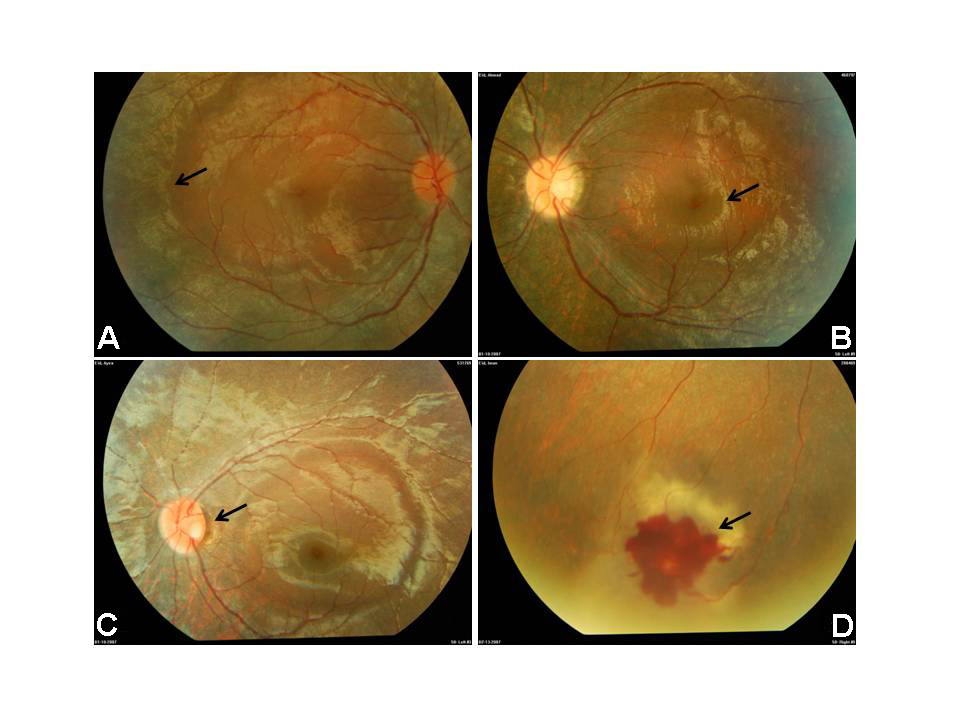
 Figure 3 of
Imtiaz, Mol Vis 2012; 18:1885-1894.
Figure 3 of
Imtiaz, Mol Vis 2012; 18:1885-1894.