Figure 4. Optical coherence tomography (OCT) of the retinal nerve fiber layers (RNFLs). A: The right eye (upper) and left eye (lower) OCT of RNFLs for II:1 show global thinning of all RNFLs. B: The right eye (upper) and left eye (lower) OCT of RNFLs for II:3 show temporal thinning of RNFLs in both eyes.
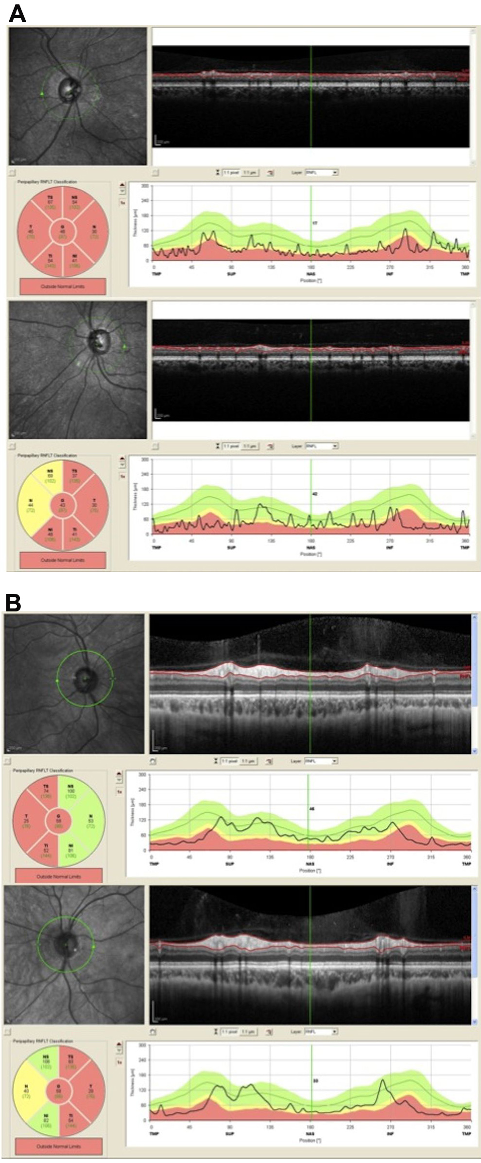
 Figure 4 of
Désir, Mol Vis 2012; 18:1849-1857.
Figure 4 of
Désir, Mol Vis 2012; 18:1849-1857.