Figure 6. Histological section of C57BL/6 mouse eye imaged with our multiphoton microscope. Eye structures appear normal after imaging.
No distortion or photocoagulation is noted in the tissues near the drainage angle of the eye. Blue dye was used to mark the
orientation of the eye before enucleation (*). S=sclera. R=retina. I=iris. C=cornea. L=lens. The scale bar represents 50 µm.
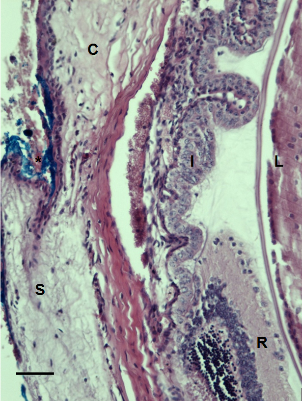
 Figure 6 of
Masihzadeh, Mol Vis 2012; 18:1840-1848.
Figure 6 of
Masihzadeh, Mol Vis 2012; 18:1840-1848.