Figure 4. Two-dimensional tiled multiphoton images of the angle of the eye taken at different depths. Distinct features of the anatomy
of the eye are visible. Images from top (A) to bottom (D) represent sections taken at different depths below the surface of the eye from (A) 580 μm, (B) 540 μm, (C) 520 μm, and (D) 500 μm. From left to right, we can see the iris (I), trabecular meshwork region (TM), sclera (S) and conjunctiva (C).
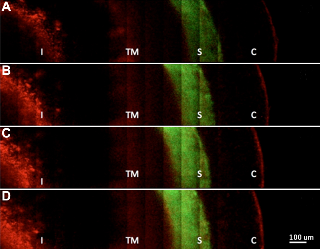
 Figure 4 of
Masihzadeh, Mol Vis 2012; 18:1840-1848.
Figure 4 of
Masihzadeh, Mol Vis 2012; 18:1840-1848.