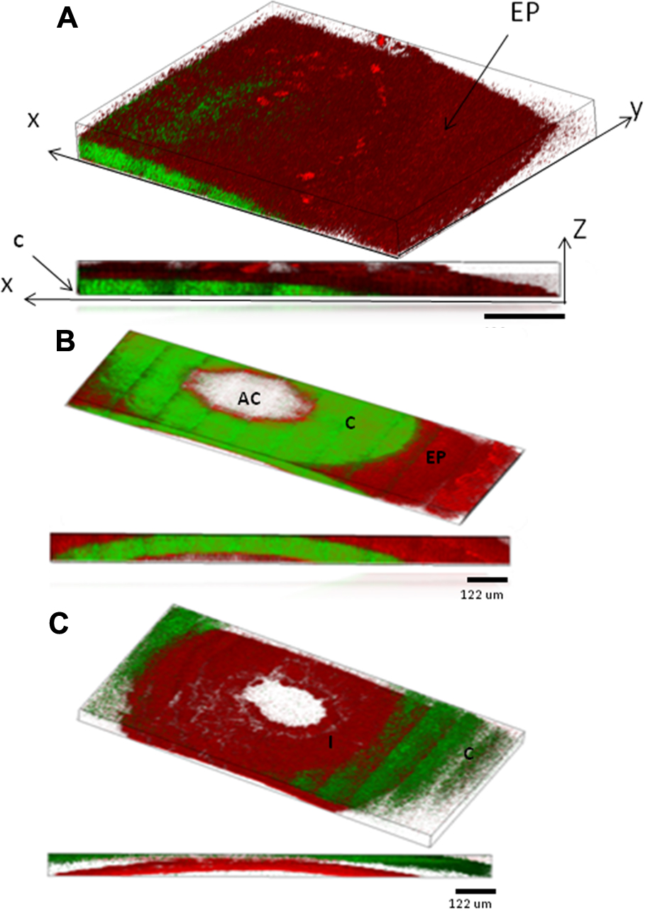
Figure 3. Three dimensional reconstructions of the images of the mouse eye taken with our multiphoton microscope. Below each 3-D reconstruction
is a two-dimensional projection showing the side view of the image composite. The cornea (C) and iris (I) are indicated on
the images.
A: Image reconstruction of one quarter of the cornea section (indicated as section A-A in
Figure 2). The epithelium layer (EP) is visible by the strong 2-photon autofluorescence signal (red; 2PAF) clearly delineating it
from the stroma of the cornea that emits predominately second-harmonic generation from collagen (green;SHG).
B: Image reconstruction of a cross section through the anterior chamber (AC; section B-B in
Figure 2). The 2PAF signal on the outer rim of the image is from the epithelial layer on top of the stroma while the SHG signal is
from the collagen in the stroma.
C: Image reconstruction of the measured region deep into the anterior chamber that includes the iris (I) which emits 2PAF signal
predominantly from the pigment granules.
 Figure 3 of
Masihzadeh, Mol Vis 2012; 18:1840-1848.
Figure 3 of
Masihzadeh, Mol Vis 2012; 18:1840-1848.