Figure 1. A schematic of the custom multiphoton microscope illustrating the beam path of the excitation laser and the emitted signals.
The pulsed laser light is first sent through a prism compressor (to ensure the shortest pulse duration at the sample) and
passed through two galvanometric scanning mirrors for raster scanning at the sample. The excitation laser beam travels through
a scan lens (SL) and tube lens (TL) before being sent into the microscope through the side port. A custom made lens relay
system is used to convert our inverted microscope (Olympus IX71) into an upright microscope for in situ imaging. The multiphoton
signal from the sample is collected back through the microscope objective and separated from the excitation laser light with
a dichroic mirror (DM1). An additional dichroic mirror (DM2) is used to separate the SHG and 2PAF signals, which are subsequently
focused on separate photo-detectors. Abbreviations: prism (Pr), half-wave plate (HW), polarizer (PP), telescoping optics (T),
objective lens (Obj). Inset shows a photograph of the custom stereotaxic device that holds the mouse and orients the eye position
and angle with respect to the objective lens. A reservoir is held over the mouse eye to keep the eye moist during imaging
sessions.
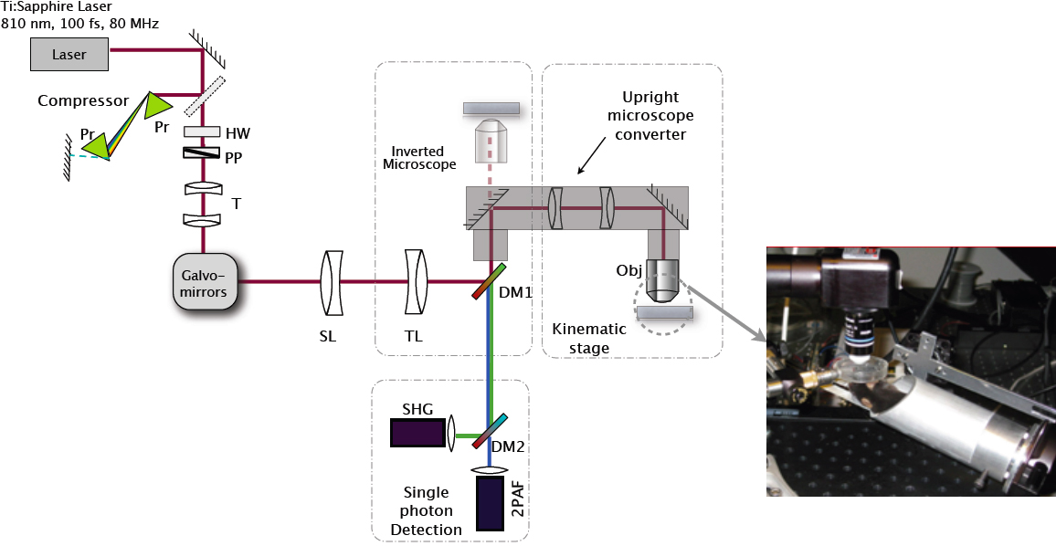
 Figure 1 of
Masihzadeh, Mol Vis 2012; 18:1840-1848.
Figure 1 of
Masihzadeh, Mol Vis 2012; 18:1840-1848.