Figure 5. Morphology of the seeded cells. Mesenchymal stem cells (MSCs; p=1) adhered to the acellular corneal matrix (ACM) after 12
h (A; 10×), and after 7 days, the seeded MSCs showed a ceroid appearance (B; 10×). A scanning electron microscopic (SEM) showed that the MSCs adhered to the ACM tightly, with masses of microvilli on
their apical surface (E; 2,000×). Corneal limbal stem cells (LSCs; p=1) adhered on the ACM after 12 h (C; 10×), and after 7 days (D; 10×). SEM showed a flat, squamous, polygonal appearance (F; 2,000×). Under the seeded microenvironment, MSCs and LSCs presented similar proliferation ability with cells under the normal
culture condition. There were no significant differences in the proliferation of the seeded MSCs and corneal LSCs as determined
with MTT (G).
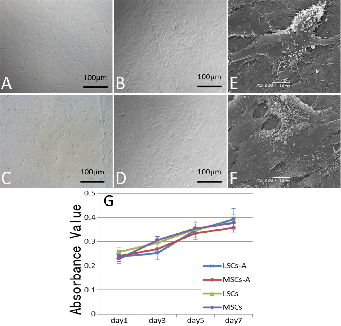
 Figure 5 of
Zhang, Mol Vis 2012; 18:161-173.
Figure 5 of
Zhang, Mol Vis 2012; 18:161-173.