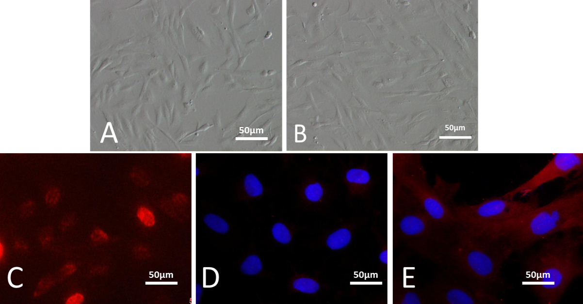
Figure 2. Limbal stem cells cultured in vitro. Inverted phase contrast microscopy showed the morphology of limbal stem cells. Cells
appeared to be compact, uniform, and cobblestone-pavement in shape (A; 20×), with 70% confluence at day 7 (B; 20×). The overwhelming majority of cells were positive for p63 (C; 20×) and ABCG2 (red; E; 20×) indicated the presence of limbal epithelial progenitor cells, only a few of which were keratinK3/12 positive (red merged
with DAPI; D; 20×).

 Figure 2 of
Zhang, Mol Vis 2012; 18:161-173.
  Figure 2 of
Zhang, Mol Vis 2012; 18:161-173.