Figure 6. Corneal scarring and keratocyte cell density in rabbit corneas 3 months after lamellar keratectomy in eyes treated with and
without MMC. Second harmonic (A and B) and confocal imaging of the same plane stained with Syto59 (C and D) of control (A and C) and 0.2 mg/ml MMC treated corneas (B and D) 2 months before lamellar keratectomy injury. Control corneas 3 months after LK injury showed the presence of irregularly
organized scar collagen (A) and a high density of scar fibroblast nuclei (B). By comparison, eyes treated with MMC 2 months before LK injury showed normal lamellar stromal collagen at the epithelial-stromal
interface (C) and markedly decreased corneal keratocytes (D) 3 months after LK injury.
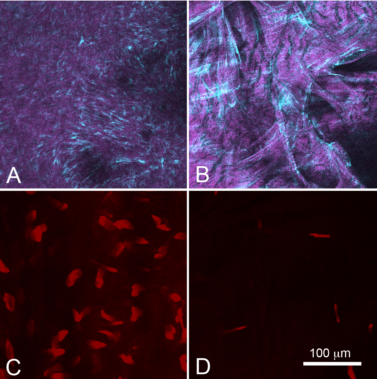
 Figure 6 of
Jester, Mol Vis 2012; 18:1828-1839.
Figure 6 of
Jester, Mol Vis 2012; 18:1828-1839.