Figure 5. In vivo confocal microscopy of lamellar keratectomy injury in rabbit corneas 2 months after MMC treatment. In vivo confocal
microscopy of rabbit corneas receiving vehicle control (A and B) or 0.2 mg/ml MMC (C and D) 2 months before lamellar keratectomy (LK). In control eyes, 3 months after LK injury, there is a dense scar underlying the
epithelium (Epi) detected in the XZ projection (A) that contains densely populated scar fibroblasts seen in the XY plane underlying the epithelium (B). Eyes treated 2 months earlier with MMC showed no scar in the XZ projection (C) and decreased keratocytes underlying the epithelium in the XY plane (D) 3 months after LK injury. Quantification of light scattering showed that eyes treated with MMC 2 months before LK injury
had significantly less haze (E).
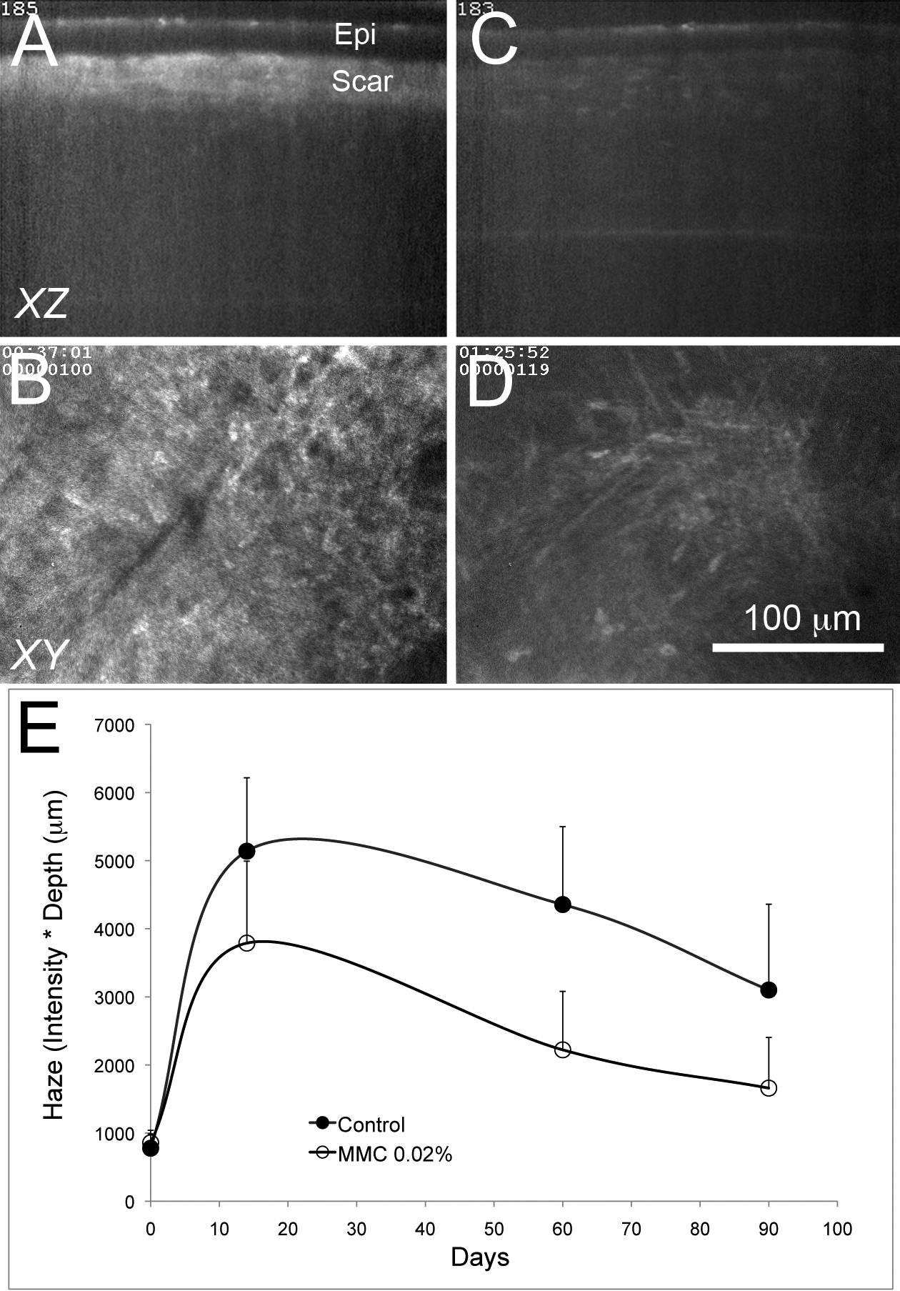
 Figure 5 of
Jester, Mol Vis 2012; 18:1828-1839.
Figure 5 of
Jester, Mol Vis 2012; 18:1828-1839.