Figure 3. DNA repair in quiescent
keratocytes compared to proliferating fibroblasts following MMC
exposure. MMC treated Keratocytes (A and B) and
corneal fibroblasts (C and D) were stained with
γH2AX (A and C, green), Ki67 (B and D,
green) and DAPI (red) 24 h after treatment. Keratocytes showed
only γH2AX staining while fibroblasts showed γH2AX and Ki67
staining. Comet assay (E) shows that MMC dose dependently
increases the Comet tail in fibroblasts 24 h after treatment,
while significantly decreasing the Comet tail in quiescent
keratocytes.
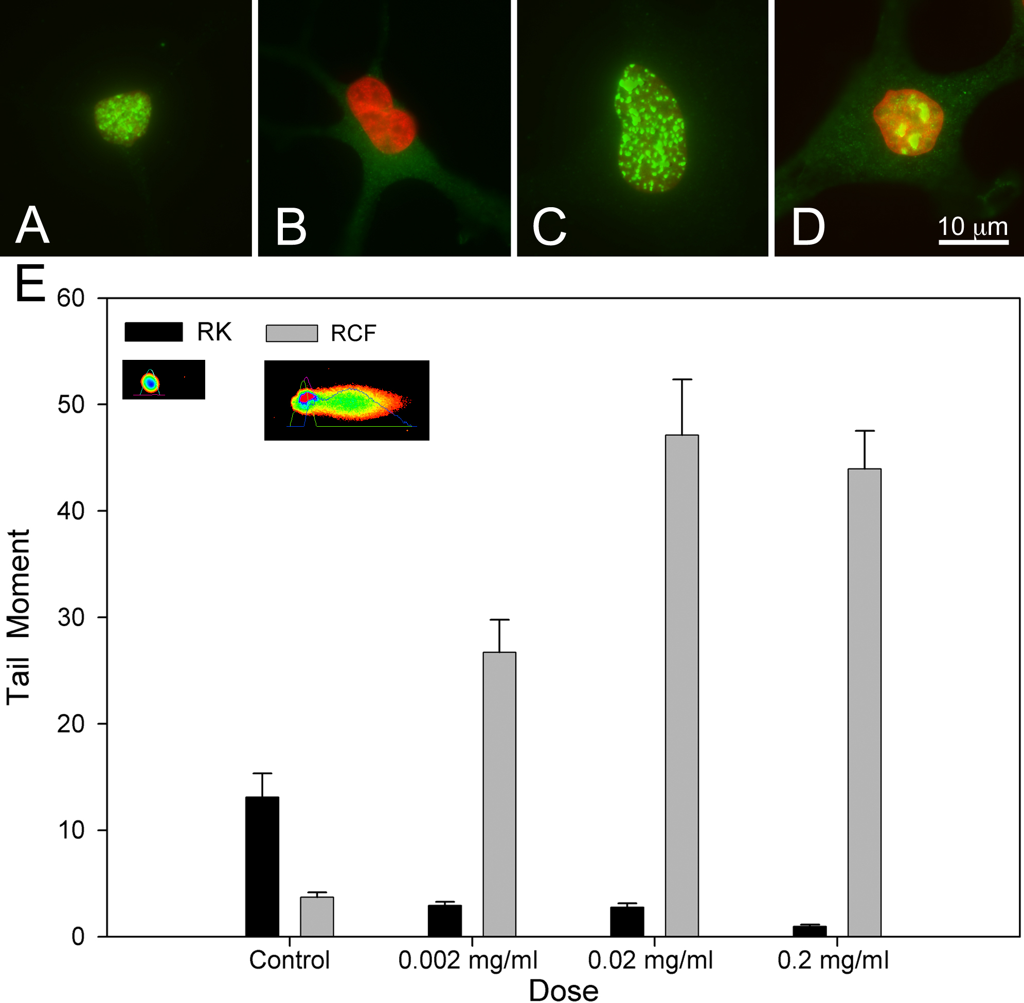
 Figure 3
of Jester, Mol Vis 2012; 18:1828-1839.
Figure 3
of Jester, Mol Vis 2012; 18:1828-1839.