Figure 1. Concentration-dependent
effects of MMC on keratocytes treated with TGFβ for 72 h. Cells
were untreated (A) or treated with TGFβ alone (B)
or after 0.02 mg/ml MMC (C) or 0.002 mg/ml MMC (D)
treatment and then stained with phalloidin (red), anti-αSMA
(green), and DAPI (blue). Cells treated with TGFβ alone showed a
spread morphology and αSMA staining typical of myofibroblasts (B),
while cells treated with MMC before TGFβ showed loss of cell
spreading and decreased αSMA staining (C and D).
Quantitation of αSMA staining (E) showed significantly
reduced numbers of myofibroblasts following pre-treatment with
MMC and decreased protein expression for αSMA (insert).
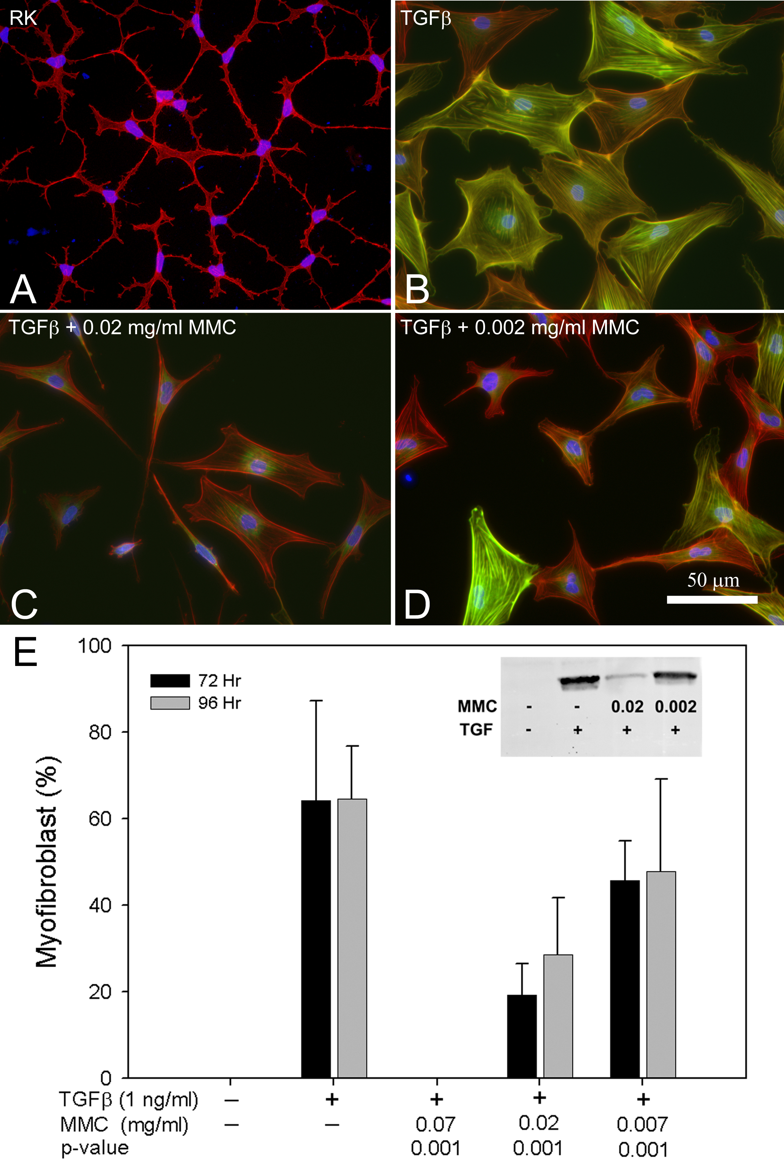
 Figure 1
of Jester, Mol Vis 2012; 18:1828-1839.
Figure 1
of Jester, Mol Vis 2012; 18:1828-1839.