Figure 7. Quantification of fluorescence intensity for IL-1β and TNF-α. Mean fluorescence intensity for IL-1β and TNF-α in different
groups was quantified and compared among them. BTX-B-injected mouse cornea and conjunctiva showed IL-1β and TNF-α staining
significantly higher than that of normal and saline-injected mice at the 1 and 2 week time points (p<0.01, t-test). FML-treated mice with BTX-B injection revealed a 66% and 60% reduction in IL-1β and TNF-α staining respectively in
the corneal and conjunctival epithelia at the 2 week time point (p<0.01, t-test). FML- fluorometholone-treated group, Nep- nepafenac-treated group, Ket- ketorolac-treated group.
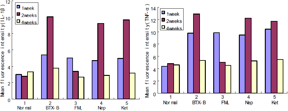
 Figure 7 of
Zhu, Mol Vis 2012; 18:1803-1812.
Figure 7 of
Zhu, Mol Vis 2012; 18:1803-1812.