Figure 5. Immunofluorescent staining with IL-1β antibody (red) in corneal epithelia (CE: arrows) and conjunctival epithelia (CJE: arrowheads)
were detected in FML-treated mice at 1 week BTX-B post-injection, decreased significantly at 2 and 4 weeks post-injection
compared to other groups (D-F and J-L with nuclear counterstain in blue). Negative and isotype controls, which did not stain, were not shown. Scale bar: 50 µm.
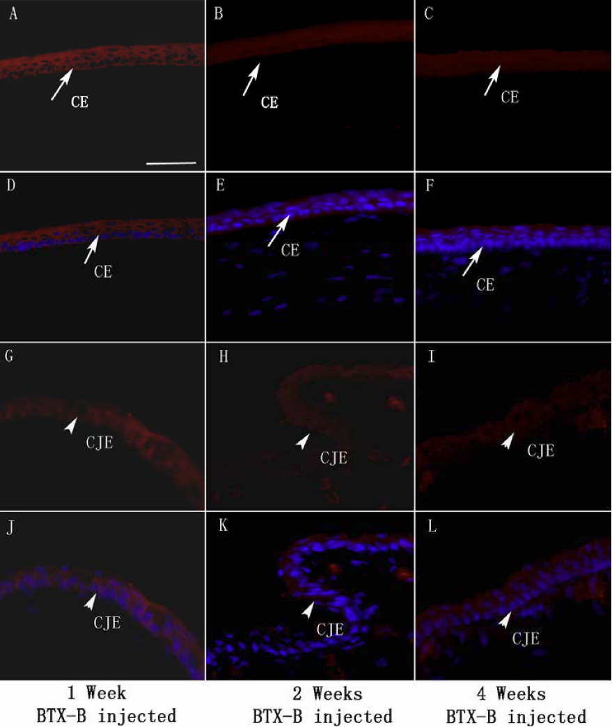
 Figure 5 of
Zhu, Mol Vis 2012; 18:1803-1812.
Figure 5 of
Zhu, Mol Vis 2012; 18:1803-1812.